What is Atelectasis?
Atelectasis is the collapse of a lung or one of the lobes within a lung. It occurs when the small air sacs in the lung, called alveoli, lose air.
Commonly seen as a respiratory complication after surgery, atelectasis can also be a complication of respiratory problems such as cystic fibrosis, chest injuries, lung tumors, respiratory weakness, and fluid accumulation in the lungs. Additionally, atelectasis can develop if a foreign object is inhaled.
In individuals with pre-existing lung disease, the occurrence of atelectasis can make breathing difficult. The treatment to be applied varies depending on the severity of the cause of the collapse.
Atelectasis Diagnosis Criteria
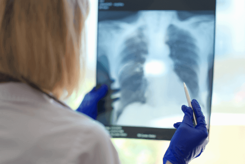
Atelectasis can be diagnosed directly through a physical examination and a chest X-ray. However, to confirm the source of the symptoms or to determine the type and severity of atelectasis, other tests may be applied. These tests include:
- Computed tomography (CT) scan
- Oximetry
- Chest ultrasound
- Bronchoscopy
Symptoms of Atelectasis
The causes of atelectasis may not always be obvious. Therefore, one of the following symptoms may occur:
- Difficulty breathing
- Coughing
- Wheezing
- Rapid, shallow breathing
- Blue skin and lips
- Chest pain
Causes of Atelectasis
Atelectasis is often seen after surgical procedures. During surgery, the use of anesthesia prevents taking deep breaths that fully inflate the lungs or coughing to clear mucus from the lungs. This can lead to alveolar blockage and lack of air, causing absorptive atelectasis. Other causes of atelectasis include:
- Mucus accumulation in individuals with cystic fibrosis, children, and severe asthma attacks after surgery
- Accidental inhalation of an object blocking the lung
- Fluid accumulation around the lungs due to heart diseases
- Air accumulation around the lungs
- Non-cancerous benign tumors
- Malignant tumors
- Scarring in the lung
- Respiratory infections such as COPD, acute respiratory distress syndrome, pneumonia, or Covid-19
- Lung infections
- Lung tissue scarring
Risk Factors for Atelectasis
Factors that increase the risk of atelectasis include:
- Any condition that makes swallowing difficult
- Smoking
- Prolonged bed rest without sufficient position changes
- Pain or injuries that make coughing painful or lead to shallow breathing, including abdominal pain or broken ribs
- Lung diseases
- Medications that cause weak breathing
- Recent abdominal or chest surgeries
- Respiratory muscle weakness due to spinal cord injury or other neuromuscular disorders
- Recent general anesthesia
Complications of Atelectasis
Atelectasis affecting a small area is usually treatable, especially in adults. However, some complications may arise due to atelectasis. These complications include:
- Low blood oxygen
- Lung inflammation
- Respiratory failure
Treatment of Atelectasis
The treatment for atelectasis varies depending on the cause. In mild cases, it may resolve on its own without treatment. In some cases, various medications are used to loosen and thin mucus. However, if the atelectasis is caused by an obstruction, surgery or other treatments may be required.
Chest Physiotherapy
Also known as chest physiotherapy, this treatment aims to clear the airway. Techniques that help take deep breaths after surgery are applied to expand the collapsed lung tissue. Learning these techniques before surgery is more effective. Techniques in chest physiotherapy include:
- Using a portable device called a spirometer to perform deep breathing exercises and then coughing to help clear the lungs. This technique can help eliminate mucus and other secretions.
- Positioning the body so that the head and chest are lower, allowing mucus to flow better into the lower parts of the lungs.
- Lightly tapping the chest area to loosen mucus. This technique is called percussion. It can be applied with a hand-held device or a high-frequency chest wall oscillation vest.
Surgery
Suctioning mucus from the lungs or performing bronchoscopy can clear airway blockages. During bronchoscopy, doctors use a flexible tube inserted through the throat to clear the airways.
If a tumor causes atelectasis, surgery to remove the tumor, as well as other treatments like chemotherapy and radiation, may be used.
Breathing Treatments
In some cases, a breathing tube may be needed. Continuous positive airway pressure (CPAP) is used after surgery for individuals who are too weak to cough and those with low blood oxygen levels, known as hypoxemia.
Types of Atelectasis
Atelectasis is divided into many subtypes, with three main types. The main types of atelectasis include:
Obstructive Atelectasis
Also known as absorptive atelectasis, obstructive atelectasis occurs when oxygen and carbon dioxide in the alveoli enter the bloodstream, and no new air enters. This causes the alveoli to collapse. Surgical procedures requiring anesthesia are a common cause of absorptive atelectasis.
Objects that block the lungs and prevent air from entering the alveoli can also cause absorptive atelectasis. This blockage can be due to mucus, tumors, or foreign objects.
Compressive Atelectasis
Also known as compressive atelectasis, it occurs when factors such as fluid, air, blood, or tumors around the lungs press on the lungs, causing them to collapse.
Contraction Atelectasis
Scarring in the lung can cause contraction atelectasis. The scar tissue prevents the alveoli from opening correctly, leading to lung collapse.
Other Types of Atelectasis
A rare type of atelectasis, known as irregular atelectasis, can be found in newborns, especially premature newborns or individuals with acute respiratory distress syndrome. Patchy atelectasis occurs when there is an insufficient amount of protein to help prevent the lungs from collapsing.
Other types of atelectasis are used to describe the area where the collapse occurs, its appearance, or its severity. These types of atelectasis include:
- Bibasilar atelectasis
- Round atelectasis
- Gravity-dependent atelectasis
- Subsegmental atelectasis
Difference Between Atelectasis and Bronchiectasis
Atelectasis occurs due to the sudden collapse of lung tissue, while bronchiectasis is the localized destruction of lung tissue due to loss of elasticity in the walls of the airways. As a result, bronchiectasis involves irreversible dilation of the airways caused by the damage.
Although both conditions lead to lung diseases, their pathologies are very different. The main difference is that atelectasis involves sudden blockage, while bronchiectasis involves gradual destruction leading to dilation.
Difference Between Atelectasis and Pneumothorax
The definition of atelectasis is broader than that of pneumothorax. In pneumothorax, air leaks into the space between the lung and the chest wall, causing part or all of the lung to collapse. Therefore, pneumothorax is one of the various causes of atelectasis.