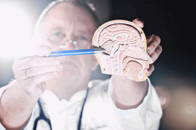
What is cerebellar herniation?
Also known as Chiari malformation, cerebellar herniation is a growth abnormality where brain tissue at the bottom of the skull extends into the spinal canal. The cause of this abnormality is structural issues such as the skull being smaller than expected.
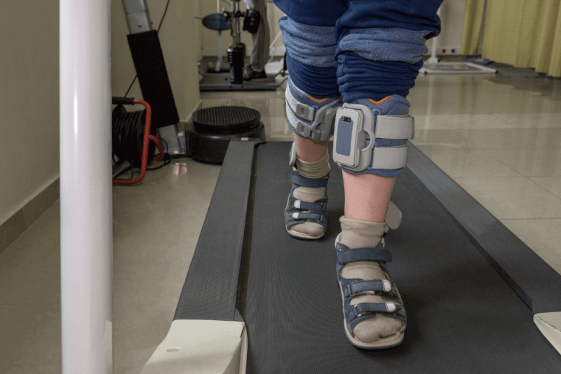
When there is not enough space in the skull, the cerebellum, in particular, grows downward through an opening at the base of the skull called the foramen magnum. The cerebellum is the part of the brain that controls muscle movements, posture, balance, speech, and coordination. Therefore, abnormalities in this structure cause symptoms such as balance loss.
Additionally, this abnormality causes pressure on the base of the skull. As a result, the flow of cerebrospinal fluid is obstructed. This fluid cushions the brain and spinal cord as much as possible, circulates nutrients and chemicals, and removes waste products.
Diagnosis criteria for cerebellar herniation
Cerebellar herniation is usually diagnosed after a full physical examination. During this examination, doctors test the patient's movements, balance, and sensations in the hands and feet.
Additionally, memory problems, learning difficulties, and developmental delays in children are considered. Various tests are used to confirm the diagnosis by obtaining a detailed image of the brain and spinal cord. These tests include:
- Magnetic resonance imaging (MRI)
- Cine MRI
- Computed tomography (CT)
- X-ray
Fetuses with cerebellar herniation can be imaged via ultrasound before birth.
Causes of cerebellar herniation
A significant portion of cerebellar herniation cases involves a structural growth abnormality in the brain or spinal cord. This causes pressure in this part of the brain. This pressure leads to the cerebellum growing in an unexpected location. These abnormalities form during fetal development.
Cerebellar herniation is seen in almost all cases at birth. Although rare, some cases may develop cerebellar herniation in later ages. The causes of cerebellar herniation in older ages include:
- Brain tumor
- Cyst
- Hematoma
- Hydrocephalus
- Intracranial hypertension
- Pseudotumor cerebri
In addition, cerebellar herniation can be seen as a complication in the following health conditions:
- Goldenhar syndrome
- Achondroplasia
- Ehlers-Danlos syndrome
- Connective tissue disorders
- Spina bifida
Symptoms of cerebellar herniation
Common symptoms of cerebellar herniation include:
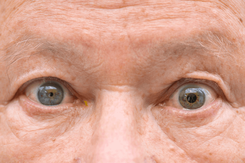
- Double vision
- Blurred vision
- Abnormal eye movements
- Light sensitivity
- Tinnitus
- Balance loss
- Scoliosis
- Loss of bladder or bowel control
- Fainting
- Sleep apnea
Symptoms in patients vary from person to person. Therefore, while some patients show no symptoms, others may experience severe symptoms.
Symptoms due to the disease are usually seen from birth. In some patients, symptoms may not appear until late childhood or adulthood. Additionally, symptoms due to cerebellar herniation may improve or worsen at different points over time.
Treatment methods for cerebellar herniation
The treatment of cerebellar herniation is planned according to the severity of the symptoms. Patients with no symptoms are usually not treated. However, the patient's condition is monitored with regular check-ups.
Patients with mild symptoms such as headache or neck pain can consider the following treatment options:
- Pain relievers to help manage symptoms
- Massage therapy
- Physical therapy
- Limiting physical activities such as heavy lifting
- Using medical devices such as hearing aids or glasses to assist with hearing or vision loss
Cerebellar herniation surgery
Surgery may be recommended for patients with severe symptoms caused by cerebellar herniation. Cerebellar herniation surgeries can be performed using different procedures, including:
Craniotomy
In this method, surgeons remove part of the skull to relieve pressure on the brain and help the flow of cerebrospinal fluid.
Posterior fossa decompression
This surgical method is the most common procedure used to treat cerebellar herniation. In this method, the surgeon removes the posterior fossa, the part of the skull's back, to relieve pressure on the brain and create more space.
Laminectomy
Depending on the severity of cerebellar herniation, the surgeon removes a small portion of the bones covering the spine to restore the flow of cerebrospinal fluid and create more space for the spinal cord.
Duraplasty
In this method, the surgeon opens the membrane outside the brain. Then, to enlarge the membrane and give the brain more space, a patch is sewn. This increases space and relieves pressure on the brain.
Electrocautery
To create more space and allow the drainage of cerebrospinal fluid, surgeons can apply a small amount of electricity to reduce a small section called the cerebellar tonsils. The cerebellar tonsils retract without causing brain or nerve damage.
Shunt placement
In this method, used for hydrocephalus patients, a shunt or tube is placed in the skull to collect more cerebrospinal fluid. This allows the fluid to flow from the baby's skull to another part of the body.
How long does cerebellar herniation surgery take?
Cerebellar herniation surgeries usually take 2 to 3 hours. After surgery, patients are taken to the intensive care unit for the first few hours to determine if any complications from brain trauma are observed.
What should cerebellar herniation patients pay attention to?
The hospital stay after cerebellar herniation surgery can last from a few days to several weeks, depending on the patient's condition. If patients are elderly or have ongoing health problems, they need to stay in the hospital longer to avoid any complications.
After being discharged, patients should limit their daily activities for a few weeks. As they recover, they can gradually return to their normal daily lives. Activities that patients should limit after surgery include:
- Bending
- Lifting heavy objects
- Straining
- Prolonged coughing periods
- Doing house or garden work
- Consuming alcoholic beverages
- Driving until approved by the doctor
It is expected for patients to feel pain or discomfort when turning or lifting their heads after surgery. Pain relievers are prescribed to alleviate this situation.
Additionally, fatigue, weakness, and headache are common after surgery. During this period, it is very important to monitor for signs of infection such as redness, inflammation, fever, or fluid accumulation around the incision.
If headaches worsen, severe neck pain occurs when the head is tilted forward, increased drowsiness, or weakness in the arms or legs, the doctor should be contacted.
In the weeks following surgery, patients gradually return to normal activities. However, it is recommended to avoid strenuous activities and exercises during this time. Instead, walking helps improve blood circulation and muscle tone during recovery.
In addition, neck pain and stiffness are common problems after surgery. To overcome these issues, it is important to do light exercises to strengthen the muscles and perform isometric neck exercises.
Types of cerebellar herniation
Cerebellar herniation can be seen in five different types.
Type 1
Type 1 cerebellar herniation occurs when the lower part of the cerebellum extends into the hole at the base of the skull. Normally, the spinal cord passes through this hole. Type 1 is the most common type of cerebellar herniation and usually does not cause symptoms. If symptoms occur, the first signs are seen during adolescence or adulthood.
Type 2
Type 2 cerebellar herniation occurs during the development of the fetus in the womb. In this form, the cerebellum and brainstem grow abnormally, causing pressure inside the skull. Type 2 is usually associated with a condition called myelomeningocele, a severe form of spina bifida. This condition occurs when the spine and spinal cord canal do not close properly before birth. After birth, the spine can be closed with surgery. However, there is a risk of paralysis for the babies.
Type 3
Type 3 is the most severe type of cerebellar herniation. It occurs when part of the cerebellum and brainstem extend outside the skull through an abnormal opening at the back of the skull. This opening is called the foramen magnum. Additionally, some membranes surrounding the brain and spinal cord extend through this opening. This results in a swelling called encephalocele at the back of the skull. Extremely severe, it appears in childhood. Symptoms in children include neurological problems, learning delays, and seizures. It is life-threatening and usually requires surgery.
Type 4
Type 4, one of the rare types of cerebellar herniation, occurs when the cerebellum is underdeveloped or some parts are missing. Extremely severe, type 4 is usually life-threatening in infants.
Type 0
Type 0 cerebellar herniation is a very rare type. In this type, there is a very small part of the cerebellum or none at all in the hole at the base of the skull. However, many symptoms appear at this level, and the reason for the symptoms is the abnormal flow of cerebrospinal fluid near the base of the skull.
Complications of cerebellar herniation
Cerebellar herniation can lead to serious health problems and threaten life. Complications that may be seen due to the disease include:
Hydrocephalus
In a life-threatening condition called hydrocephalus, cerebrospinal fluid accumulates in the brain. Accumulation of cerebrospinal fluid causes pressure inside the skull. This can lead to problems with cognitive functions and an irregularly shaped skull. If left untreated, it is life-threatening. This complication is frequently seen in type 2.
Syringomyelia and Hydromyelia
When cerebrospinal fluid does not flow properly between the brain and spine, it can accumulate in the spine. This fluid accumulation can damage the spinal cord. Additionally, it can cause symptoms such as movement and balance problems, pain, muscle weakness, muscle spasms, numbness, decreased sensitivity to heat and cold, and loss of bladder and bowel control.
Tethered cord syndrome
Children born with myelomeningocele are at risk of developing tethered cord syndrome as they grow. This condition occurs when the spinal cord sticks to the spine due to a scar from the closure surgery. In tethered cord syndrome, slow and progressive nerve damage is seen, affecting the lower body and leg muscles. Additionally, bowel and bladder functions can be affected.
People who experience insomnia or severe headaches due to the disease may also experience mood changes. This can lead to the development of depression.