30-Second Summary
- Clubfoot, one of the birth defects typically present at birth, is used to describe a variety of foot abnormalities in which the shape or position of the foot is distorted.
- Clubfoot can range from mild to severe. In about half of the babies born with this issue, the deformity affects both feet. Babies born with clubfoot may face difficulty walking normally as they grow older.
- The exact cause of clubfoot is unknown. However, it is believed to result from a combination of genetic and environmental factors.
- Clubfoot is typically diagnosed through ultrasound imaging performed during pregnancy.
What is Clubfoot (Pes Equinovarus)?
Clubfoot, one of the birth defects typically present at birth, is used to describe a variety of foot abnormalities in which the shape or position of the foot is distorted. In clubfoot cases, the tissues connecting muscles to bones are abnormally shorter. For babies born healthy, clubfoot is usually an isolated condition.
Clubfoot can be mild or severe. In about half of the babies born with this condition, the deformity affects both feet. Babies born with clubfoot may face difficulty walking normally as they grow older. Therefore, doctors usually recommend treatment right after birth.
Surgical procedures are generally not needed for clubfoot treatment. However, some babies may require surgery to walk healthily.
What Causes Clubfoot (Pes Equinovarus)?
The exact cause of clubfoot is still unknown. However, it is believed to be a combination of genetic and environmental factors.
Symptoms of Clubfoot (Pes Equinovarus)
Possible symptoms of clubfoot in babies include:
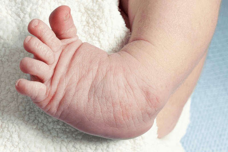
- The top of the foot is usually bent downward and inward, causing the arch to increase and the heel to rotate inward.
- The foot may be so severely turned that it appears to be upside down.
- The affected leg or foot may be slightly shorter than the other.
- The calf muscles of the affected leg are usually underdeveloped.
- Despite the appearance of the foot, it does not usually cause pain.
- Stiffness in the ankle.
- Limited range of motion in the foot.
Additionally, the following changes in foot shape may be observed:
- The foot may appear kidney-shaped.
- Deep wrinkles may be seen on the inside of the foot.
- The foot arch may be higher than normal.
Diagnosis Criteria for Clubfoot (Pes Equinovarus)
Clubfoot is typically diagnosed through ultrasound imaging performed during pregnancy. The treatment plan for clubfoot is usually formulated during this period.
In some cases, the diagnosis is made after birth, usually during the baby’s first physical examination. In some cases, an X-ray may be required to confirm the diagnosis.
Treatment Methods for Clubfoot (Pes Equinovarus)
Due to the extreme flexibility of newborns' bones, joints, and tendons, treatment begins in the first one or two weeks after birth. The goal of the treatment is to improve the functioning of the feet before the child learns to walk in order to prevent long-term disability.
Ponseti Method for Clubfoot Treatment
The Ponseti method is the most commonly used treatment for clubfoot. The stages of this treatment include:
- Initially, the doctor places the foot in a cast to maintain its correct position.
- Every few weeks, the foot is re-casted to reposition it properly over several months.
- At the end of the process, a surgical procedure is performed to lengthen the Achilles tendon.
After the foot shape is corrected, the following measures are taken to maintain the foot position:
- Stretching exercises for the baby’s foot.
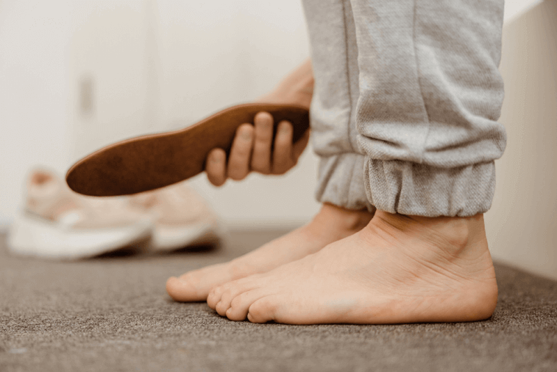
- Use of special shoes and supports.
- The shoes and supports should be worn 24 hours a day for approximately three years.
For this method to be successful and prevent the foot from returning to its previous shape, the foot supports must be used according to the doctor’s instructions. The main reason for failure in this method is usually the improper use of the supports.
French Method for Clubfoot Treatment
The French method, similar to the Ponseti method, uses splints and taping instead of casting. The splint is a device that helps support and protect the bones.
This treatment, performed by a physiotherapist, should start shortly after birth. The splint and taping need to be used every day. The physiotherapists teach the parents how to apply them. Several physiotherapy appointments are required each week.
The steps in the French technique include:
- The baby’s foot is stretched into the correct position.
- The foot is secured using tape and a splint.
- The procedure is repeated every day for two months.
- Afterwards, the frequency of the treatment is reduced until the baby reaches 3 months old.
In babies treated with the French method, Achilles tendonectomy is also required. By the end of the three months, improvements in the clubfoot can be seen. However, to maintain the correct foot position, the protective treatment must continue until the child reaches 2-3 years of age.
Surgery for Clubfoot
If non-surgical options do not yield positive results or if the condition is severe, surgical procedures may be considered. Performed by orthopedic surgeons, this operation involves lengthening and repositioning the tendons and ligaments to bring the foot into a better position. After surgery, a cast is usually applied for two months, and a support is used for about a year.
Despite surgery, there is still a possibility that the clubfoot will not be fully corrected. However, in most cases, babies who undergo surgery can wear regular shoes and lead an active life.
Surgical Methods for Clubfoot
Reconstructive surgery, which reshapes the foot, is typically used in clubfoot surgery. The surgeon may reshape and reposition a bone in the leg or foot.
In addition, external fixation may be used to achieve the reshaping. In this surgical method, pins and wires are used to apply external supports to the foot. These supports are adjusted daily to gradually reposition the foot.
Benefits of Clubfoot Surgery
Clubfoot surgery helps bring the baby’s feet to a normal position when other methods are unsuccessful. After these procedures, the success rate is high, and the child can walk normally.
Complications of Clubfoot Surgery
Complications following clubfoot surgery may vary depending on the procedure used. However, common complications include:
- Damage to the foot nerves
- Swelling of the feet
- Problems with blood flow to the foot
- Stiffness
- Arthritis
- Weakness
Clubfoot (Pes Equinovarus) Risks
Clubfoot typically does not cause complications until the baby reaches walking age. If treated, normal walking is usually achieved. However, some challenges may arise in the following areas:
- The affected foot may be less flexible than the other foot.
- The affected leg may be slightly shorter, but this usually does not cause significant mobility issues.
- The affected foot may be 1-1.5 shoe sizes smaller than the other foot.
- The calf muscles of the affected leg may be weaker than those of the other leg.
If untreated, clubfoot can lead to more serious problems, including:
- Development of arthritis in the child.
- The unusual appearance of the foot may cause body image concerns during adolescence.
- Ankle sprains may prevent the child from walking properly, causing them to walk on the outer side or top of the foot.
- Changes in walking can hinder the natural growth of the calf muscles.
- Large wounds and calluses may form on the foot.
- Abnormal walking patterns may develop.
How to Help a Child with Foot Support?
Using supports according to the instructions is crucial for improving the success rate. However, it can be difficult for children to wear supports properly. The following tips can help ease this process:
- Play with the child and do light exercises while wearing the supports.
- After the first three months, the supports are only used during naps and nighttime sleep, making it easier to incorporate them into the child’s sleep routine.
- Adding soft padding to metal bars on the supports.
- Avoid using creams or lotions while wearing the supports.
- Ensure the shoe does not slip off the support.
Types of Clubfoot
There are two types of clubfoot:
Isolated or Idiopathic Clubfoot
This is the most common type of clubfoot. In this case, the child has no other medical issues. Idiopathic means the cause is unknown.
Non-Isolated Clubfoot
This type of clubfoot occurs along with other health problems, including joint issues and spina bifida. Neural tube defects are related to problems with the baby’s brain, spine, and spinal cord.