What is keratoconus?
Keratoconus, one of the eye diseases, is a condition in which the cornea becomes thinner and bulges outward in the shape of a cone. The cone-shaped cornea causes blurred vision and makes patients more sensitive to light and glare. Keratoconus, which usually affects both eyes, causes one eye to be affected more than the other in some cases. Keratoconus, which especially affects people between the end of adolescence and the age of 30, is a disease that progresses slowly over 10 years.
If keratoconus is diagnosed in the early stages, vision problems can be corrected with glasses or soft contact lenses. In the later stages, different types of lenses such as rigid gas permeable contact lenses or scleral lenses are required. If the condition worsens, corneal transplantation is recommended.
A procedure called collagen cross-linking can help slow and stop the progression of keratoconus. In this way, the need for cornea transplantation in patients can be avoided.
Keratoconus diagnosis
For keratoconus, the patient's medical history is first examined. An eye examination is then performed. Tests that can be performed to learn more about the shape of the cornea include
- Light refraction in the eye is tested with a special device called a phoropter. In this device, lenses with different lens numbers are placed in front of the patient's eyes, and then the lens with the sharpest vision is determined by the patient.
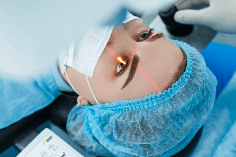
- In a slit lamp examination, a beam of light is sent perpendicular to the surface of the eye. A low-power microscope is then used to visualize the eye under this light. With this test, the ophthalmologist evaluates the shape of the cornea and looks for other possible problems in the eye.
- Keratometry, which involves focusing a circle of light on the cornea, allows the basic shape of the cornea to be determined.
- In computerized corneal mapping, detailed mapping of the cornea is performed with corneal tomography and topography. The thickness of the cornea can also be measured during this test. This test allows early diagnosis of keratoconus, usually before a slit lamp examination.
Causes of keratoconus
Genetic and environmental factors are thought to play a role in the causes of the disease. Studies have shown that one in 10 patients has a parent with the disease. Risk factors that can cause the disease include the following.
- Retinitis pigmentosa
- Down syndrome
- Marfan syndrome
- Ehlers danlos syndrome
- Hay fever
- Asthma
- Severe formation of the eyes
- Family history of keratoconus
Keratoconus symptoms
The symptoms caused by the disease vary in its later stages. Among the symptoms seen in the early stages are the following.
- Blurred or distorted vision
- Sudden worsening or blurred vision
- Problems with night vision
- The need for frequent changes in eyeglass prescriptions
- Increased sensitivity to bright light or glare
Complications of keratoconus
In some cases, the cornea swells rapidly, causing a severe reduction in vision. This also causes scarring of the cornea. The breakdown of the inner lining of the cornea plays a role in this condition. The formation of this condition called descement membrane causes fluid to enter the horn. For this reason, the swelling usually goes down spontaneously, but there may be scarring that affects vision.
Progression of the disease can lead to corneal injury. Injury to the cornea leads to worsening vision problems and the need for corneal transplantation.
Keratoconus treatment methods
Keratoconus treatment varies depending on the severity of the disease. Basically, there are two different approaches to treating the disease. The main goals of these treatments are to improve vision and slow the progression of the disease.
Keratoconus surgery
It is a preferred method in the presence of a scar on the cornea. In addition, surgery is recommended in cases of excessive thinning of the cornea or difficulty in seeing with strong lenses. The surgery performed varies according to the swelling of the cornea.
Keratoconus surgery methods
Surgeries are known by different methods according to the needs of the patients. These methods include the following.
Intrastromal corneal ring segments
ICRS is a procedure for patients with mild to moderate keratoconus. In this method, small synthetic rings are placed on the cornea. These rings help to flatten the cornea. Flattening of the cornea can also improve vision and help contact lenses fit better. In some cases, this procedure is performed in combination with corneal cross-linking.
Cross-linking
Especially in patients with keratoconus due to aging, cross-linking surgeries are performed to strengthen the collagen structure in the cornea. This operation takes approximately half an hour for each eye. The aim of the surgery is to make the collagen structure of the cornea much more resistant. In this way, it is aimed to prevent corneal thinning and swelling.
Cornea transplant
Patients need corneal transplantation in cases of scarring or excessive thinning. Depending on the situation, specialists may replace all or part of the cornea with healthy donor tissue. Corneal transplantation is also called keratoplasty.
Keratoconus surgery benefits
The most important advantage of surgery for patients is that it stops the progression of the disease and eliminates visual impairment. In this way, it is possible to significantly improve the quality of life of patients.
Keratoconus surgery complications
Corneal transplantation in keratoconus patients is a treatment method with generally successful results. However, as with other surgical procedures, there are some complications that may occur in corneal transplantation. These complications include the following.
- Graft rejection
- Poor vision
- Infection
- Astigmatism
- Glaucoma
- In the treatment of astigmatism complication after corneal transplantation, the problem is usually eliminated by using hard contact lenses.
Complications that may occur after corneal cross-link surgery include the following.
- Dryness in the eyes
- Eye pain or irritation
- Further worsening of the infectious disease
Complications that can be seen in patients after corneal ring segments surgery include the following.
- Infection
- Corneal thinning
- Glare or the appearance of halos of light
Life after keratoconus surgery
After their surgery, the life of the liner changes positively. Surgeries prevent further progression of the disease and prevent complete loss of vision. However, these surgeries do not correct existing visual impairments. For this reason, patients are required to wear special lenses or glasses produced in the appropriate size for their subject after surgery.
The healing process of keratoconus patients
The healing process after keratoconus treatment varies according to the surgical method. Patients may complain of pain for 1 to 2 days after cross linking treatment, and these complaints may be accompanied by light sensitivity. A one-week rest period is required for the patient to return to work or school. It is also recommended that patients use drops for one month after surgery. After cross linking surgery, patients are advised to attend follow-up appointments every 6 months until the age of 40. In addition, it is most important that the patient does not rub his/her eyes.
The most important point is that patients should not rub their eyes in the early period after this eye surgery performed with Femtosecond laser, which does not require hospitalization. After surgery, patients are advised to wait 2-3 days before taking a shower. Patients are also advised to take medication for one month after surgery. Usually 2-3 days is enough time for them to return to their daily lives.
The first 2 days after laser treatment may feel painful. Painkillers or cold complex application is recommended for pain relief. A period of 8-10 days is required for patients to return to their daily lives. During this period, patients are advised to avoid exposure to sunlight and wear sunglasses for the next 3 months. The time required for patients to reach full vision is one month. Laser treatment, which is usually applied together with cross linking treatment, is a lifelong treatment. Therefore, no further deterioration is expected.
What should individuals with keratoconus pay attention to?
There are 2 points that keratoconus patients should pay attention to. The first is not to rub their eyes. In addition, it is important to have regular check-ups after diagnosis and to keep the progression of the disease under control.
Keratoconus lenses
The most important criterion used to determine kerata contact lenses is the stage of the disease. Therefore, in order to determine the correct lens, corneal capture and keratometric reagent values should be used together to determine the appropriate lens for the patient. There are 4 different lens types recommended for keratoconus patients
Gas permeable keratoconus lenses
These lenses, also called hard lenses, have a hard structure when touched. These lenses can perfectly pass oxygen to the cornea, but they are difficult to use and have the risk of falling out. For this reason, it can be extremely difficult to play sports with gas permeable lenses.
Gas permeable lenses mask the irregular corneal surface caused by the disease and provide a smooth surface. It is a type of lens used to improve the vision of patients with second and third stage keratoconus. These lenses, which are placed at the sharpest point of the cornea, can be of different diameters.
Soft keratoconus lenses
Soft keratoconus lenses, which offer comfortable lens wear, have an extremely high moisture retention capacity. Since these lenses cause less friction on the cornea, eye irritations are prevented. It is preferred by many patients because it provides moisture while efficiently delivering the oxygen required for the cornea. However, they are not sufficient in terms of image quality in advanced stages. Since they need to be replaced every 3 months, their cost is higher than gas permeable lenses, but they are much more comfortable to wear because they are soft.
Hybrid keratoconus lenses
These lenses, which are soft around the periphery and hard in the middle, have been used by patients since the early 2000s. The center surfaces of these lenses are made of gas permeable hard lens material, while the edges are made of silicone or polyurethane soft lens material. Although it offers a much more comfortable use than gas permeable hard lenses, various problems may arise because oxygen transmission does not occur at the same level in the soft part.
Although the cornea can obtain the oxygen it needs, the moisture problem is eliminated in these lenses. In addition, the use of 2 different materials in the same lens also causes handling problems. For this reason, patients with hybrid keratoconus lenses need to use artificial tear drops regularly.
Scleral keratoconus lenses
These lenses, which have very high oxygen permeability, move much less with the eye due to their large size. For this reason, it is one of the most comfortable keratoconus lenses. Since it is not made of a soft material, it is possible to use it for a long time. In terms of visual quality, it is the type of lens that provides the best result.
All lens types mentioned above are specially prepared for patients. For this reason, the measurement values made by the doctor are transmitted to the factories and then production is made on behalf of the patient. Since this process, which takes approximately one month, takes place in factories abroad, the costs are calculated in foreign currency.
Keratoconus laser
Laser and radiation treatments are used to stop the progression of keratoconus and improve vision. In particular, cross linking is the recommended treatment for patients up to the age of 35. This treatment is a radiation therapy to stop the progression of the disease. For this reason, it is not included among surgical procedures. The success rate of cross linking treatment at the right time is very high.
No touch laser treatment
Options such as no touch laser treatment used in the treatment of keratoconus are laser options that allow patients with low-numbered eye disorders to see more clearly without glasses. Although it varies depending on the thickness of the cornea, to touch laser treatment, which is applied to myopic astigmatism degrees up to 3 to 3.5 numbers, is also known as hybrid laser.
PTK Laser treatment
PTK laser treatment is also a laser treatment applied together with cross linking treatment. It is applied to patients with unclear vision, including with glasses. With this laser treatment, the corneal surface is made more regular. This allows patients to see more clearly with glasses.
Keratoconus stages
Keratoconus disease is divided into 3 stages as early, middle and advanced depending on the changes seen in the cornea
Phase 1
In the first stage, which is the early stage, patients have no symptoms, but if diagnosed, the lenses to be used can be soft or hard. In addition, patients are known to be nearsighted and glasses against astigmatism are also known to be recommended.
Phase 2
In the second stage, corneal shape changes become evident. There is also corneal thinning. Cross-linking treatment is usually used to prevent keratoconus progression from this stage. Laser treatment can also be used to improve the quality of vision and reduce astigmatism.
Phase 3
The third stage is the most advanced stage of the disease. Although corneal thinning is severe in patients at this stage, scarring can also be observed. In the treatment of patients at this stage, especially corneal ring segments and cornea transplantation are possible. However, these treatments are rarely needed in patients undergoing cross-linking treatment in the second stage.