30-Second Summary
- Kienböck's disease refers to the degradation of the lunate bone, which is a crescent-shaped bone in the wrist. This degradation can occur over several years or within a few months.
- The diagnosis of Kienböck's disease is generally made through physical examination. During the examination, providing as specific information as possible about the pain location and complaints is crucial.
- Surgical methods are typically preferred for treating Kienböck's disease. The procedure to be applied depends on the stage of the disease.
- The stage of Kienböck's disease can be determined through MR and CT scans. These imaging systems help identify blood flow and fractures.
What is Kienböck's Disease?
Kienböck's disease refers to the degradation of the lunate bone, a crescent-shaped bone in the wrist. The degradation of the bone can happen over several years or within a few months. The degradation worsens over time, and symptoms become more severe. In some cases, it may also cause arthritis in the wrist.
Not everyone with Kienböck's disease shows symptoms. Therefore, in some cases, the disease is discovered during the examination of the wrist for a different health problem.
What Causes Kienböck's Disease?
The exact cause of Kienböck's disease is unknown, but it is believed that several factors contribute to the bone degradation. These risks include:
- Reduced blood flow to the lunate bone, which is the most commonly accepted cause among experts. Insufficient nourishment of the bone can lead to its death.
- Trauma resulting in damage to the lunate bone.
- Problems in the wrist can occur when the ulna and radius bones of the forearm are not of equal length.
- The shape of the lunate bone may be congenitally abnormal.
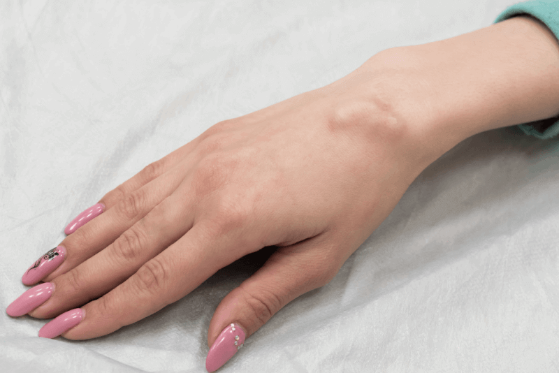
Symptoms of Kienböck's Disease
Symptoms that may occur in individuals with Kienböck's disease include the following:
- Pain similar to that felt when the wrist is sprained.
- Swelling of the wrist.
- Stiffness in the wrist.
- Weakness in the wrist.
- Crepitus or crackling sounds when moving the wrist.
- Reduced range of motion in the wrist.
- Reduced strength in the wrist.
Diagnosis Criteria for Kienböck's Disease
Kienböck's disease is usually diagnosed through physical examination. During the examination, it is important to provide specific information about the location of the pain and the complaints. Additionally, the following tests may be required to confirm the diagnosis:
- X-ray
- MRI
- CT scan
- Bone scan
Treatment Methods for Kienböck's Disease
Although there is no definitive cure for Kienböck's disease, the following measures can help manage the condition:
- Restoring blood flow to the lunate bone.
- Pain relief.
- Improvement of wrist mobility.
- Stopping the progression of Kienböck's disease.
Non-Surgical Treatments for Kienböck's Disease
Non-surgical treatment options for Kienböck's disease include:
- Immobilizing the wrist in a cast for several months to allow more blood to flow to the lunate bone.
- Occupational therapy to reduce pain and teach how to properly use the wrist.
- Corticosteroid injections and over-the-counter pain relievers such as ibuprofen and naproxen.
Kienböck's Disease Surgery
Surgical methods are typically preferred for treating Kienböck's disease. The procedure used depends on the stage of the disease.
Surgical Methods for Kienböck's Disease
There are five primary surgical procedures used for the treatment of Kienböck's disease. These procedures include:
Joint Leveling
If the two bones in the forearm (radius and ulna) are of unequal lengths, joint leveling may be necessary. This is because the unequal bone lengths put extra pressure on the lunate bone, causing it to collapse. In this procedure, a small piece of bone may be removed from the radius or ulna, or a bone graft may be applied.
Revascularization
This procedure aims to restore blood flow to the lunate. In revascularization, a small piece of bone is taken from another part of the body and placed in the lunate. Pins are used to keep the bones together while they heal. This procedure is only performed in stages 1 and 2 of the disease.
Fusion
Partial fusion or full fusion can be performed. In this permanent procedure, the surgeon fuses some or all of the bones in the wrist together. If full fusion is performed, the wrist will not be able to move, although the forearm may still be rotated.
Implant Arthroplasty
In this procedure, the lunate bone is replaced with a prosthesis.
Proximal Row Carpectomy (PRC)
This procedure involves the removal of the lunate bone along with other carpal bones in the wrist if the lunate is broken.
Benefits of Kienböck's Disease Surgery
Surgical procedures for Kienböck's disease help relieve symptoms caused by the condition. The benefits of the surgery vary depending on the procedure used. Some procedures improve blood flow, helping to stop the progression of the disease, while others prevent further complications.
Complications of Kienböck's Disease Surgery
The complications following Kienböck's disease surgery can vary based on the procedure performed. However, some general complications may include:
- Anesthesia-related complications
- Infection
- Bleeding
- Damage to wrist nerves and blood vessels
Stages of Kienböck's Disease
The stage of Kienböck's disease can be determined through MRI and CT scans. These imaging systems help understand blood flow and identify fractures. The stages and characteristics of the disease are as follows:
Stage 1
In the first stage, patients feel pain similar to that of a sprained wrist. The cause of this pain is not clear but is generally believed to be due to decreased blood flow to the lunate.
Stage 2
Due to the lack of blood flow to the lunate, the bone begins to harden. This hardening process is called sclerosis, and it indicates that the bone is dying.
Stage 3
In this stage, the lunate bone begins to break. The broken bones may cause the other bones to move, which leads to more pain. Additionally, it becomes difficult to firmly grip objects, and the range of motion is limited.
Stage 4
In the final stage of the disease, the outer parts of the bones near the lunate become weakened. At this stage, arthritis in the wrist may be observed.