30-Second Summary
- Osteopetrosis is a rare disease that causes bone tissue to become abnormally dense.
- In addition to being rare, the densification of bones also makes them more prone to fractures.
- Osteopetrosis is divided into three subtypes. The reason for the classification is the differences in genetic inheritance.
- The only currently effective treatment is stem cell therapy. This treatment is applied to the malignant infantile type using hematopoietic stem cell transplantation.
What is Osteopetrosis?
Osteopetrosis is a disease that causes bone tissue to become abnormally dense. In addition to being one of the rare diseases, the densification of bones also makes them more prone to fractures. It is characterized by increased bone density due to a defect in bone resorption by cells called osteoclasts.
This condition leads to the accumulation of bone with defective architecture. The accumulation of defective bone can lead to various skeletal abnormalities and make bones more susceptible to fractures. Osteopetrosis is also referred to as marble bone disease, Albers-Schönberg disease, and osteosclerosis fragilis generalisata.
Types of Osteopetrosis
Osteopetrosis is divided into three subtypes. The reason for the classification is the differences in genetic inheritance. Types of osteopetrosis include:
Autosomal Dominant Osteopetrosis (ADO)
Also known as Albers-Schönberg disease, autosomal dominant osteopetrosis (ADO) is the mildest form among the types. For this reason, some patients do not show any symptoms. As a result, overly dense bones are diagnosed incidentally during X-rays taken for the investigation of another disease.
This common form is inherited as recessive and X-linked recessive. In individuals who show symptoms, initial signs may appear at different times from late childhood to adulthood.
Autosomal Recessive Osteopetrosis (ARO)
This type, which is more severe than ADO, presents symptoms from birth. Individuals with this type may suffer bone fractures from seemingly insignificant bumps and falls. The skull bones of these individuals may also be excessively dense, which can compress the nerves in the head and face.
This can lead to vision and hearing loss and facial paralysis. Additionally, due to the density of bone structure, the function of the bone marrow may be impaired. This may disrupt the system responsible for producing new blood and immune cells.
As a result, individuals with severe ARO often experience anemia, severe bleeding, and recurrent infections. Also known as malignant infantile type, this form typically shortens life expectancy.
Intermediate Autosomal Osteopetrosis (IAO)
The rarest form of osteopetrosis, IAO has been observed in only a few individuals to date. The symptoms of this type usually appear during childhood. Therefore, the risk of bone fractures and anemia increases after this period. However, life-threatening bone marrow abnormalities are not observed. Additionally, some individuals have shown abnormal calcium accumulation in the brain. As a result, intellectual disability and a type of kidney disease known as renal tubular acidosis may occur.
Causes of Osteopetrosis
Mutations in at least eight genes can cause osteopetrosis. Various mutations in the gene called CLCN7 are responsible for approximately 75% of autosomal dominant osteopetrosis cases and 10-15% of recessive osteopetrosis cases.
Mutations in the TCIRG1 gene are seen in approximately 50% of autosomal recessive osteopetrosis cases. Mutations in other genes are less common causes of the autosomal dominant and autosomal recessive forms of the disorder. In about 30% of all cases, the cause is unknown.
All genes that cause osteopetrosis are responsible for the formation, development, and function of special cells called osteoclasts. The role of these cells is to break down old bone tissue and allow the formation of new bone tissue, shaping the bones. This is the most critical process for maintaining healthy bone tissue.
Mutations in these genes cause abnormal or deficient osteoclasts. The absence of functional osteoclasts leads to a situation where old bone tissue is not broken down even though new bone tissue is formed. As a result, bones become excessively dense.
Symptoms of Osteopetrosis
The characteristic symptom of osteopetrosis is the excessive density of bone tissue. Other symptoms vary depending on the type of the disease. These symptoms include:
ARO
Symptoms caused by ARO appear from birth. Without immediate treatment, the life expectancy is at most 10 years. Symptoms vary depending on the type of gene mutation. Symptoms seen in the most severe type of osteopetrosis, the malignant infantile type, include:
- Abnormal bone hardening
- Increased fragility of ribs and long bones
- Large head and small jaw structure
- Growth retardation causing nerve compression
- Increased density of cranial bones
- Obstruction of normal cerebrospinal fluid flow
- Abnormal enlargement of cerebral ventricles, causing fluid accumulation
- Increased brain pressure due to hydrocephalus
- Weakening of the retina
- Protrusion of the eyes from the orbit
- Strabismus
- Involuntary rhythmic eye movements
- Blindness
- Reduction in bone marrow cavity
- Significant deficiency in all blood cells
- Blood cell production outside of the bone marrow
- Excessive enlargement of the spleen and liver due to extramedullary hematopoiesis
- Formation of myeloid tissue in the mentioned organs
- Pneumonia and frequent urinary tract infections
- Rhinitis
- Anemia
- Hematological problems appearing before neurological problems
- Hearing loss
- Eating disorders
- Difficulty in acquiring motor skills that require coordination
- Delayed tooth development
- Severe tooth decay
- Low calcium levels in the blood
- Seizures due to low calcium levels in the blood
- Rare but severe neurodegeneration
ADO Symptoms
Since symptoms usually appear in late childhood or adulthood, this type is also called the adult type. Possible symptoms include:
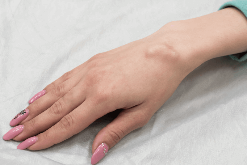
- Bone fractures after minimal trauma
- Dental abscess
- Osteomyelitis, especially in the jaw
- Cranial hyperostosis
- Rhinitis
- Hepatosplenomegaly
- Anemia
- Extramedullary hematopoiesis
Intermediate Autosomal Symptoms
This type, a combination of autosomal recessive and dominant forms, usually starts showing symptoms in childhood. The severity of symptoms varies depending on whether ARO or ADO traits are more dominant. Although the severity may vary, symptoms differ from classical recessive and dominant forms. These symptoms include:
- Abnormal bone hardening
- Fractures
- Mandibular osteomyelitis
- Knock knees with distinctly separated ankles
- Cranial hyperostosis
- Gradual deterioration of optic nerves
- Vision loss
- Muscle weakness
- Rhinitis
- Protrusion of the lower jaw
- Retention of primary teeth
- Tooth crown malformation
- Tooth decay
- Facial paralysis
- Hepatosplenomegaly
- Anemia
- Reduced platelet count in the bloodstream
- Pancytopenia
- Extramedullary hematopoiesis
AIO Symptoms
This extremely rare type presents very severe symptoms. In addition to the typical signs of osteopetrosis, symptoms include:
- Immunodeficiency
- Localized fluid retention
- Tissue swelling
- Abnormalities in hair, skin, nails, and sweat glands
Osteopetrosis Diagnosis Criteria
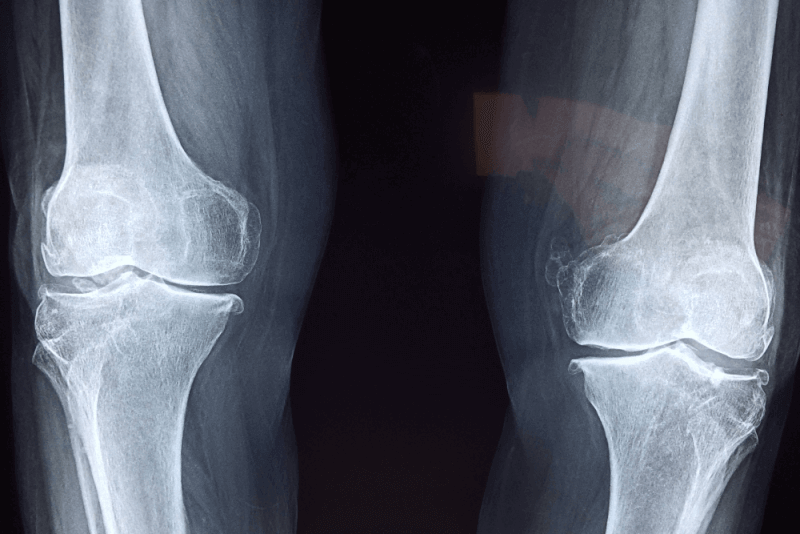
In the diagnosis phase of osteopetrosis, in addition to a detailed clinical evaluation, various special tests such as the patient's medical history, X-ray imaging, and measurement of increased bone density are performed. Since X-ray findings are highly specific, they are considered sufficient for diagnosis. Biochemical findings such as increased creatine kinase BB isoenzyme and elevated tartrate-resistant acid phosphatase concentrations also help confirm the diagnosis.
Osteopetrosis Treatment Methods
Currently, the only effectively applied treatment is stem cell therapy. In this treatment applied to the malignant infantile type, patients receive hematopoietic stem cell transplantation. This transplant enables the restoration of bone resorption by osteoclasts. Genetic studies are required to determine whether this treatment method is suitable.
Because in some gene mutations, the treatment is ineffective. In all mutations found in the OSTM1 gene and in some mutations in the CLCN7 gene, progressive neurodegeneration occurs, and stem cell therapy is unresponsive.
In mild cases of osteopetrosis, the benefits and risks of stem cell therapy must be weighed before proceeding. This is because side effects such as stem cell rejection, severe infections, and high blood calcium levels causing high mortality in the first year may be more serious than the symptoms caused by the disease.
Additionally, the following treatment options may be considered:
- Patients for whom stem cell therapy is not suitable may receive corticosteroid treatment. However, there is insufficient evidence to support its routine use in practice.
- Interferon gamma-1b can delay disease progression in individuals with severe malignant infantile osteopetrosis.
Moreover, the patients’ nutrition is very important. Calcium and vitamin D supplements should be taken to increase low calcium levels in the blood. Other treatments aim to alleviate symptoms and provide supportive care for the patient.