What is Pterygium?
Pterygium is a triangular-shaped, pink, fleshy tissue growth on the cornea. It usually starts from the inner corner of the eye and grows towards the pupil, but in some cases, it can start from the outer part of the eye and grow towards the pupil.
Although it may look alarming, it is not dangerous. The slow-growing tissue can stop at a certain point. However, it can cover the pupil completely, causing vision problems. Pterygium can affect one or both eyes.
While it usually doesn't cause serious problems, it can give the feeling that something is in the eye. It can also cause redness and irritation. In such cases, surgical treatment options are recommended.
Diagnosis of Pterygium
Pterygium is diagnosed during eye examinations using a standard slit lamp microscope. Other tests that can be applied to patients include:
- Visual acuity test
- Corneal topography
Causes of Pterygium
Pterygium is thought to be caused by the excessive growth of conjunctival tissue for two different reasons. The first and most common cause is prolonged exposure to UV light. Additionally, eye irritation due to hot and dry air, wind, and dust can lead to the formation of pterygium.
Symptoms of Pterygium
Pterygium often does not cause any symptoms before it appears. When symptoms do occur, their severity can range from mild to severe. Early symptoms of pterygium include:
- A small, raised pink growth on the eye
- Red, irritated, or swollen eyes
- Dry, burning, or itchy eyes
- A sensation of a foreign object in the eye
- Tearing
Late symptoms of pterygium include:
- Increase in lesion size
- Unattractive appearance due to the size of the lesion
- Blurred or double vision
Treatment Methods for Pterygium
If the symptoms of pterygium cause discomfort, a treatment plan is needed. If vision is not affected or there is no discomfort, no treatment is necessary. However, in cases where there is itching and burning, the following treatment methods can be used:
- Over-the-counter eye ointments or artificial tears to lubricate the eye
- Steroid eye drops or ointments to reduce pain, redness, itching, and swelling
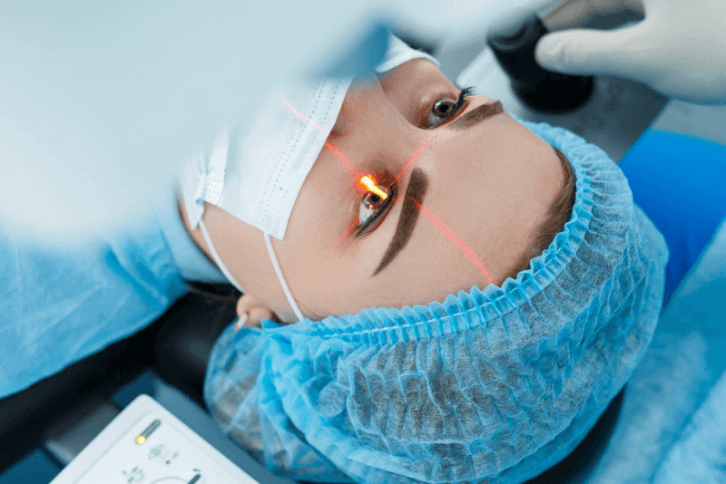
Pterygium Surgery
Surgery may be recommended if prescribed eye drops or ointments do not alleviate the symptoms or if the growth of the pterygium obstructs vision. Several methods are used in pterygium surgery, including:
- Simple removal of the pterygium
- Removal of the pterygium and placement of an amniotic membrane layer over the affected area to aid healing
- Removal of the pterygium and covering the affected area with a healthy piece of conjunctiva
Local anesthesia is applied during pterygium surgery, along with mild sedation. The pterygium is carefully removed from the eye.
In amniotic membrane surgeries, the membrane is cut to the appropriate size and placed over the area where the pterygium was removed. The membrane is then held in place with special glue or stitches.
In another method, the surgeon removes a section of conjunctiva from under the eyelid to cover the exposed area. This section is then secured in place with special glue or stitches. Pterygium surgeries typically last between 30 minutes to an hour.
Risks of Pterygium Surgery
The risks of pterygium surgery include:
- Recurrence of the pterygium. To prevent this, regular use of prescribed steroid drops and avoiding sun exposure are recommended.
- Development of cysts or infections
- Persistent double vision requiring further surgery
- Ongoing dryness and irritation in the eye
- Scleral or corneal melting