30-Second Summary
- Spina bifida, one of the most common birth defects, is caused by the incomplete development of the fetus' spine during the first months of pregnancy.
- The exact causes behind this condition are unclear. However, experts believe that a combination of genetic and environmental factors is the main cause.
- These tests do not provide definitive results. Therefore, even if blood tests show positive results, there is still a possibility that the baby will not be born with this condition.
- Treatment for spina bifida depends on the severity of the condition observed in the baby. Spina bifida occulta typically does not require treatment. However, other types may need medical intervention.
What is Spina Bifida (Split Spine Disease)?
Birth defects, one of the most common conditions, spina bifida occurs when the fetus' spine fails to develop properly during the first months of pregnancy. For this reason, it is also called a split spine.
This condition, occurring during the first 28 days of pregnancy, can be clearly observed at birth. Spina bifida, a type of neural tube defect, may not cause any health issues in some cases, but it can lead to serious symptoms in others.
In babies without visible symptoms, no treatment is required. However, babies born with severe spina bifida have open lesions on their spine, nerves, and spinal cord, which may require surgical intervention. While the lesion can be repaired surgically, nerve damage cannot be reversed, leading to permanent disability, particularly affecting the lower part of the body.
Types of Spina Bifida (Split Spine Disease)
There are three subtypes of spina bifida. These include:
Spina Bifida Occulta
Also known as hidden spina bifida, this is the most common form. In this type, one or more vertebrae (bones of the spine) may have a small separation or gap. It usually does not show any symptoms, and many people with this condition are unaware of it. It is often discovered incidentally during tests conducted for other reasons.
In rare cases, during puberty, issues may arise as the spinal cord is stretched. This could lead to symptoms such as weakness, numbness in the legs, urinary infections, and loss of control over the bladder and bowel. The severity of the symptoms corresponds to the extent of spinal cord stretching. In some cases, symptoms become severe, and surgery may be recommended.
Meningocele
A rare form of spina bifida, meningocele occurs when a fluid-filled sac protrudes through an opening in the spinal cord. No nerve tissue is affected in this case. However, babies with meningocele may experience minor functional problems with the intestines and bladder.
The sac formed by the protruding spinal cord is visible on the back. Babies with this condition can generally undergo surgery without nerve damage if the surgery is performed early in life.
Myelomeningocele
Myelomeningocele is the most severe form of spina bifida, occurring in about 1 in every 1000 births. In this form, the spinal cord does not develop properly. Part of the underdeveloped spinal cord protrudes from the back, surrounded by a sac containing brain spinal fluid and blood vessels. This sac is typically not covered by skin, and the nerves and tissues are exposed.
Around 70-90% of babies born with myelomeningocele will have hydrocephalus due to a defect at the base of the skull. If left untreated, hydrocephalus can lead to brain damage, seizures, or blindness. A shunt is placed to drain excess cerebrospinal fluid and prevent further damage.
Babies born with this form of spina bifida typically experience paralysis and weakness below the affected area. The condition can also affect the limbs, bladder, and bowel. In severe cases, even the upper body can be affected.
Spina Bifida in Adults
Adults with spina bifida face different issues than children. These problems include:
- Loss of muscle strength and flexibility, reduced physical endurance, and sensory loss appear more rapidly and severely in adults with spina bifida compared to normal aging.
- Spinal cord tethering due to the surrounding tissue causes skin wounds, rapidly progressing scoliosis, sensory loss, pain, urinary tract infections, and urinary incontinence.
- Changes in bowel function such as constipation or abdominal pain.
- Orthopedic problems like osteoporosis, early-onset arthritis, and progressive back pain.
- Skin issues such as sensory loss, circulation problems, slow wound healing, decreased sweating ability, and bruising.
- Latex allergy.
- High blood pressure.
- Obstructive and central sleep apnea that can cause long-term heart damage.
- Higher obesity rates among individuals with spina bifida.
- Women with spina bifida can become pregnant, but the pregnancy process may be more complicated.
What Causes Spina Bifida (Split Spine Disease)?
The exact causes of this condition are unclear. However, experts believe that the combination of genetic and environmental factors is the main cause. Neural tube defects, a family history of spina bifida, and inadequate intake of folate (Vitamin B9) during pregnancy are considered contributing factors.
Additionally, factors that may increase the risk of spina bifida include:
- Folate, the natural form of Vitamin B9, is extremely important for fetal development. Folic acid, a synthetic form of folate, is found in supplements and fortified foods. A very low folate level increases the risk of neural tube defects.
- Having a child with a neural tube defect increases the risk of spina bifida in subsequent pregnancies. The risk is higher if the first two children are affected. If the mother has a history of neural tube defects, the risk also increases.
- Use of anti-seizure medications such as valproic acid during pregnancy can increase the risk of spina bifida. These drugs may interfere with the body’s ability to use folate and folic acid.
- Uncontrolled diabetes in the mother before pregnancy can also increase the risk.
- Obesity during pregnancy increases the risk of spina bifida.
- Some evidence suggests that increased body temperature during the first few weeks of pregnancy, caused by factors like sauna use, hot tubs, or fever, can increase the risk of spina bifida.
Symptoms of Spina Bifida (Split Spine Disease)
Symptoms of spina bifida vary depending on the type and severity of the condition.
Spina Bifida Occulta
This type typically shows no symptoms because the spinal cord and nerves are not affected. However, some babies may have changes in the skin over the affected vertebra, such as hair growth, a pit, or a birthmark. These skin changes can sometimes be a sign of an underlying spinal cord problem, which can be diagnosed through MRI or spinal ultrasound.
Meningocele
This type may cause issues with bowel and bladder control.
Meningomyelocele
In the most severe form of spina bifida, there is an opening in the lower spine or middle back, exposing the spinal cord, tissues, and nerves. This condition can cause weakness and loss of function in the legs, as well as issues with the bladder and bowel. Hydrocephalus, which causes fluid buildup in the brain, may also occur in these babies.
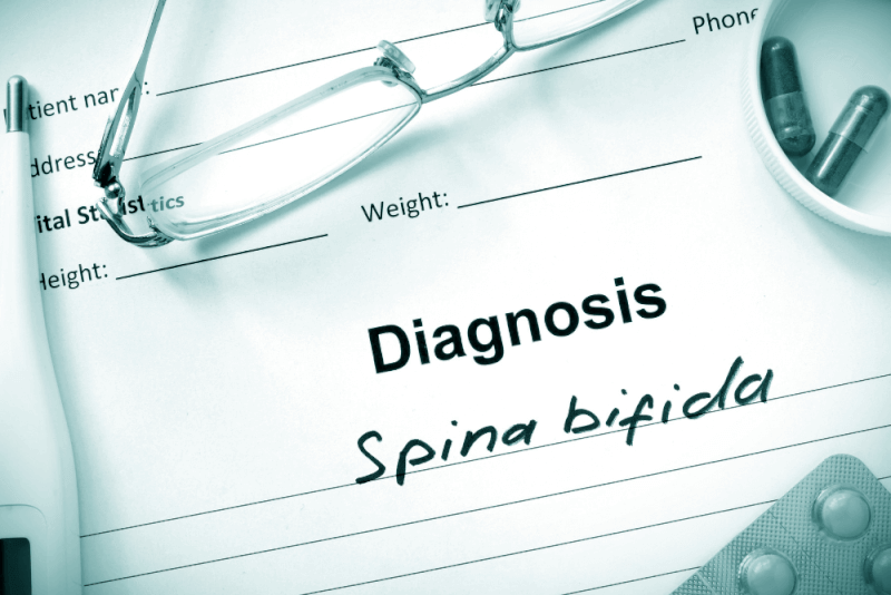
Diagnosis of Spina Bifida (Split Spine Disease)
During prenatal care, various assessments are made to check for spina bifida. However, these tests do not provide definitive results. Therefore, even if blood test results are positive, there is still a chance that the baby may not be born with this condition.
If the tests come back negative, the likelihood of the baby being born with spina bifida is very low. It is important to consult with the obstetrician to discuss the test results. Tests used to diagnose spina bifida include:
Blood Tests
Spina bifida can be detected through blood tests during pregnancy, but diagnosis is usually confirmed through ultrasound imaging.
Maternal Serum Alpha-Fetoprotein (MSAFP) Test
A blood sample is taken, and the levels of alpha-fetoprotein are tested. This protein is produced by the baby. A small amount of AFP crosses the placenta into the mother’s bloodstream, but a high level of AFP is considered a sign that the baby may have spina bifida. However, high levels of AFP are not always present in babies with this condition.
Confirmatory Test for High AFP Levels
AFP levels can vary due to factors like the baby’s age or the presence of multiple babies. Follow-up blood tests are needed to confirm high AFP levels. If levels remain high, further evaluations, including ultrasound, are required.
Other Blood Tests
Doctors may perform the MSAFP test along with two or three other blood tests to screen for other conditions, such as Down syndrome (Trisomy 21). These tests are usually conducted together with the MSAFP test.
Ultrasound
Ultrasound is the most accurate method to diagnose spina bifida before birth. It can be performed as early as the 11th to 14th week of pregnancy, but it can also be conducted between the 18th and 22nd week. Ultrasound helps identify potential birth defects that may be present at birth.
A detailed ultrasound can reveal signs of spina bifida, such as an open spine or abnormalities in the baby’s brain. In some cases, ultrasound images can show the severity of the condition.
Amniocentesis
If spina bifida is detected by ultrasound, amniocentesis may be recommended. This test involves extracting a sample of amniotic fluid from around the baby using a needle. It can help rule out other genetic disorders.
The risks of amniocentesis should be discussed with a healthcare provider, although the risk of miscarriage is very low.
Treatment for Spina Bifida (Split Spine Disease)
Treatment for spina bifida depends on the severity of the condition. Spina bifida occulta generally does not require treatment, but other forms may require medical intervention.
Spina Bifida Surgery
If spina bifida is detected in the womb, surgery can be performed both before and after birth.
Pre-birth Surgery
In babies with spina bifida, untreated nerve function can worsen. For this reason, fetal surgery is necessary to address spina bifida before birth. This surgery is typically performed before the 26th week of pregnancy.
In these surgeries, the fetal spine is exposed by opening the mother’s abdomen and uterus. The spinal cord is then repaired. In some cases, a special instrument called a fetoscope is used for a less invasive procedure.
Studies have shown that fetal surgery can reduce disability, allowing the children to require fewer mobility aids. Fetal surgery also helps reduce the risk of hydrocephalus.
However, due to the risks associated with fetal surgery, the mother and baby’s conditions must be thoroughly evaluated beforehand. Additionally, these surgeries should be performed by specialized fetal surgeons, and the team must include a fetal surgeon, pediatric neurosurgeon, maternal fetal medicine specialist, fetal cardiologist, and neonatologist.
Cesarean Section
Many babies with myelomeningocele tend to be in the breech position (with the buttocks first), making a cesarean section a safer option.
Post-birth Surgery
Babies born with myelomeningocele need surgery within the first 72 hours to close the opening in the spine. Early surgery reduces the risk of infection and further spinal cord damage.
During the procedure, neurosurgeons place the spinal cord and exposed tissue back into the baby’s body, covering it with muscle and skin. In some cases, a shunt is inserted to manage hydrocephalus.
Complication Treatment
Babies with myelomeningocele often experience irreversible nerve damage, and continuous care from doctors, surgeons, and therapists is required. Some babies may also need additional surgeries to manage complications. These treatments typically begin right after birth.
Mobility Aids
Some babies may begin exercises to help them walk with support or crutches as they grow. Mobility aids, combined with regular physical therapy, can help the child achieve independence. Some children may need walkers or wheelchairs. Children who require wheelchairs are taught how to become self-sufficient.
Bowel and Bladder Management
Routine evaluations and management of bowel and bladder function are important to reduce organ damage and disease risks. These evaluations may include x-rays, kidney scans, ultrasounds, blood tests, and bladder function tests. In the first few years of life, these evaluations are performed more frequently. As the child grows, the frequency of tests decreases. Specialists with experience in treating spina bifida can offer the most effective management options.
Bowel management can involve oral medications, suppositories, enemas, surgery, or a combination of these methods. Bladder management may include medications, catheters for bladder emptying, surgery, or a combination of treatments.
Hydrocephalus Surgery
Children with myelomeningocele may need surgery to insert a tube (ventricular shunt) to allow the fluid in the brain to drain into another part of the body. This surgery is typically performed right after birth during the procedure to close the back defect. In some cases, the shunt is inserted after the fluid buildup starts in the brain.
In some cases, a less invasive procedure called endoscopic third ventriculostomy can be an option for certain babies. However, certain criteria must be met before this procedure can be applied. During the procedure, the surgeon uses a small camera to visualize the brain and creates a hole beneath or between the brain cavities to allow cerebrospinal fluid to drain from the brain.
Other Complications Treatment and Management
Special equipment such as bathing chairs, standing frames, and other devices help improve daily living. Many complications from spina bifida can be treated to enhance quality of life. These treatments can address orthopedic issues, digestive complications, skin problems, and more.
Continuing Care
Children with spina bifida require close monitoring and care throughout their lives. Doctors assess the child’s growth, vaccination needs, and general medical issues. This care is coordinated among a team of medical specialists.
Children with spina bifida typically require ongoing treatment and continuous care from the following specialists:
- Physical medicine and rehabilitation
- Neurology
- Neurosurgery
- Urology
- Orthopedics
- Physical therapy
- Occupational therapy
- Special education
- Social work
- Nutrition
Parents and other caregivers play a vital role as part of the care team. They need to learn how to help manage the child’s condition, offer emotional and social support, and provide necessary encouragement.
What Problems Can Spina Bifida (Split Spine Disease) Cause?
Complications caused by spina bifida can range from severe to mild, affecting the individual’s quality of life. The extent of the issues depends on several factors:
- The size and location of the spinal opening
- Whether the affected area is covered by skin
- Whether any spinal nerves are exposed in the affected area
Although children with spina bifida may experience a range of complications, not all will experience all of them. Some complications can be treated, but others may persist. Common complications in spina bifida include:
Walking and Mobility Problems
Nerves controlling the leg muscles may not function properly, leading to weakness or loss of movement in the legs. This can result in paralysis or difficulty walking, depending on the location of the spinal defect and the severity of the nerve damage. Early care and therapy can have a significant impact on a child’s ability to walk.
Orthopedic Complications
Children with myelomeningocele often experience orthopedic complications such as scoliosis, clubfoot, hip dislocation, and bone and joint problems due to weak muscles. The severity of these issues depends on the position of the spinal defect.
- Scoliosis
- Clubfoot
- Hip dislocation
- Bone and joint disorders
- Muscle contractures (shortening and tightness of muscles)
Bowel and Bladder Symptoms
The nerves controlling the bowel and bladder often do not function properly in children with myelomeningocele. As a result, they may not be able to control their bowel and bladder functions.
Hydrocephalus
Babies born with myelomeningocele often experience fluid buildup in the brain, known as hydrocephalus.
Shunt Malfunction
Shunts used to treat hydrocephalus can sometimes fail or become infected, leading to changes in symptoms. Common signs of shunt malfunction include:
- Headaches
- Vomiting
- Drowsiness
- Irritability
- Swelling or redness along the shunt
- Confusion and altered consciousness
- Changes in eye movements (fixed downward gaze)
- Feeding problems
- Seizures
Chiari Malformation Type II
Chiari Malformation type II is a common condition in children with myelomeningocele. The brainstem, which is the lowest part of the brain, extends and moves lower than normal in the spinal canal or neck area. This can cause arm weakness, difficulty breathing, and swallowing problems. Rarely, this condition can lead to brain compression, requiring surgery to relieve the pressure.
Meningitis
Children with myelomeningocele are at risk for developing infections in the membranes surrounding the brain. This potentially dangerous infection can lead to brain damage.
tethered Spinal Cord
When the spinal cord becomes attached to surrounding tissue, it can lead to problems as the child grows. This attachment can cause loss of bowel and bladder control, which can be managed with surgery to limit disability.
Sleep Apnea
Sleep apnea or other sleep disorders can affect children and adults with spina bifida, particularly those with myelomeningocele. It may be necessary to perform tests to determine if there are instances of short periods of breathing cessation during sleep. Treatment can improve the quality of life for these patients.
Skin Problems
Children with spina bifida may experience reduced sensation in certain parts of their bodies. As a result, sores can form on the feet, legs, hips, and back. These wounds can become deep and infected, especially in children with myelomeningocele.
Latex Allergy
Children with spina bifida have an increased risk of developing allergic reactions to latex products. Symptoms of latex allergy include rashes, sneezing, itching, watery eyes, and runny nose. In severe cases, anaphylactic shock, which can cause swelling of the face and airway and difficulty breathing, may occur. Therefore, non-latex gloves should be used during childbirth.
Other Complications
As children with spina bifida grow, other issues may arise. These complications can include urinary tract infections, gastrointestinal issues, and depression. Children with myelomeningocele may also experience learning difficulties, such as challenges with focusing, learning math, and reading.
Prevention of Spina Bifida (Split Spine Disease)
Folic acid supplementation can reduce the risk of spina bifida and other neural tube defects. It is recommended to start taking folic acid supplements one month before conception and continue through the first trimester of pregnancy.
Folic Acid Supplementation
Having adequate folic acid in the body during the early weeks of pregnancy is essential to prevent spina bifida. However, many women do not know they are pregnant during this critical period. Therefore, it is recommended that women of childbearing age take 400 mcg of folic acid daily.
Foods fortified with folic acid, such as enriched bread, pasta, rice, and some breakfast cereals, are also beneficial. Folate, the natural form of folic acid, is also found in various foods.
- Enriched bread
- Pasta
- Rice
- Some breakfast cereals
Folic acid is often listed as "folate" on food labels. Folate is the natural form of folic acid found in foods.
Planning a Pregnancy
Women who are planning to become pregnant or could become pregnant should take 400 to 800 mcg of folic acid daily. The body does not process folate as easily as folic acid, so it is not always possible to use all the folate from foods.
For the prevention of spina bifida, it is important to take vitamin supplements. In addition to preventing spina bifida, folic acid also helps reduce the risk of other birth defects, such as cleft lip, cleft palate, and some heart defects.
A healthy diet with folate-rich foods or foods fortified with folic acid is also recommended. Some of these foods include:
- Beans
- Peas
- Citrus fruits
- Fruit juices
- Egg yolks
- Cow’s milk
- Avocados
- Dark green vegetables
Higher Doses of Folic Acid
If a mother has spina bifida or has had a child with spina bifida, the risk of having another child with the condition is increased. In this case, higher doses of folic acid are required. Additionally, women with diabetes or who take seizure medications may also need higher doses of folic acid.