What is Sturge-Weber Syndrome?
Sturge-Weber Syndrome is a rare disease and does not occur acquired. This congenital disease occurs when blood vessels in the brain and face are affected. In general, it affects areas such as the eyes, skin, mouth, jaw and nervous system.
There are three main features of Sturge-Weber Syndrome. There is a red or pink birthmark called port wine, a brain abnormality called leptomeningeal angioma and increased pressure in the eye. The manifestation of these features may vary and not all three features may be present in Sturge-Weber syndrome.
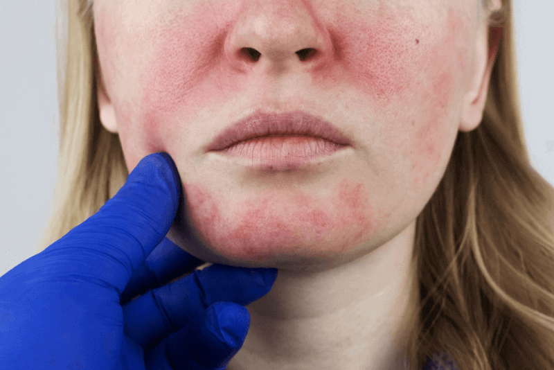
In the syndrome, the port wine birthmark is common. This type of birthmark is caused by dilated capillaries close to the surface of the skin. A port wine birthmark can usually be found on the face, typically on the forehead, temple or eyelid. Over time, the skin inside the port wine birthmark may darken and thicken.
In addition to the port wine birthmark, the two thin layers of tissue covering the brain and spinal cord show abnormal formation and enlargement of blood vessels. This anomaly, called leptomeningeal angioma, affects one or both sides of the brain, disrupting blood flow in the brain. It can lead to loss of brain tissue and calcium deposits in the brain. This condition in the brain can cause stroke-like attacks, which usually start at the age of 2. Some individuals have attention deficit hyperactivity disorder, focusing problems and learning difficulties.
Diagnostic Criteria for Sturge-Weber Syndrome
Usually the first diagnostic criterion for Sturge-Weber Syndrome is the recognition and examination of a port wine birthmark. Babies born with birthmarks may also have tests to check for problems with their brain and eyes. Some of the tests are as follows:
- MRI of the brain with or without contrast,
- A CT scan of the brain can detect excessive calcium deposits,
- EEG is checked in children with seizures,
- A detailed eye examination including pressure measurements
When diagnosing Sturge-Weber syndrome, the congenital port wine stain must first be seen. In cases where the person's brain is affected, brain scans should be performed to detect problems such as seizures, temporary muscle loss in one part of the body, learning difficulties or hyperactivity. In individuals with the syndrome, eye pressure can increase, leading to visual impairment in infancy or early adulthood. Eye pressure can increase too much in some babies, causing the pupils to dilate and swell. Nodes of abnormal-looking blood vessels can be found in various parts of the eye.
Symptoms of Sturge-Weber Syndrome
- The most prominent feature of Sturge-Weber syndrome is the port wine birthmark. The port wine birthmark can appear red or pink on the face, forehead or temples.
- The port wine birthmark is caused by dilated blood vessels near the surface of the skin and usually appears on one side of the face.
- Blood vessels in the brain and spinal cord can also enlarge and damage the brain. By disrupting the blood supply to the brain, it can lead to loss of brain tissue and calcification of the brain.
- Stroke-like attacks due to damage to blood vessels in the brain,
- Visual disturbances,
- Migraine headaches,
- Muscle weakness on one side of the body.
- People with Sturge-Weber syndrome may have normal intelligence, but in some cases intellectual disability can also occur.
- Increased pressure inside the eye, called glaucoma,
- Very dilated and swollen pupils,
- Hemangiomas in different parts of the eye,
- As the hemangioma progresses, vision loss occurs. Eye abnormalities occur on the same side of the head as the port wine birthmark.
Causes of Sturge-Weber Syndrome
Sturge-Weber syndrome is a disease caused by a mutation in the GNAQ gene. This syndrome is not inherited and the mutation is somatic, meaning that the development of blood vessels begins to be impaired in the period after conception.
The GNAQ gene mutation that causes Sturge-Weber syndrome produces a protein whose function is impaired. The protein produced fails to regulate signaling pathways and disrupts blood vessel development, causing disease.
Sturge-Weber Syndrome Treatment Methods
The treatment of Sturge-Weber syndrome varies according to the symptoms seen in the person. It can be listed as follows:
- If the patient has seizures, anticonvulsant drugs are used to reduce seizure activity,
- If eye pressure is increased, eye drops that reduce eye pressure are used,
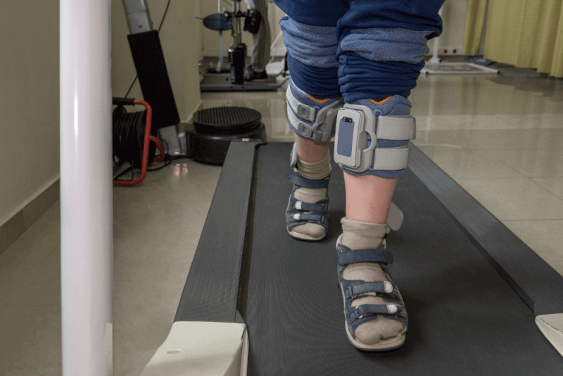
- If there are weaknesses in the muscles of the body, physical therapy can be used to strengthen the weak muscles,
- If there are problems such as mental retardation and focusing problems, special education therapies can be used,
- Laser skin resurfacing treatments are preferred to reduce the appearance of port wine birthmark.
Sturge-Weber Syndrome sclerotherapy
Sclerotherapy is a method used in the treatment of congenital vascular tangles and varicose veins. The aim is to chemically disrupt the inner surface of the vein by injecting drugs into the vascular tangle, i.e. hemangioma, which is present in Sturge-Weber syndrome, with the sclerotherapy method, turning it into a simple burn tissue.
Sturge-Weber Syndrome in Adults
Although rare in adults, Sturge-Weber syndrome can be recognized. This congenital syndrome may not be recognized in infancy and the person may continue to live with attacks until adulthood.
A person may not have a port wine birthmark on their face, and even if they do, in some cases it may go unnoticed. Sturge-Weber syndrome is diagnosed with MR imaging as a result of attacks with convulsions that occur in later life and continue throughout adulthood.
Sturge-Weber Syndrome Eye Findings
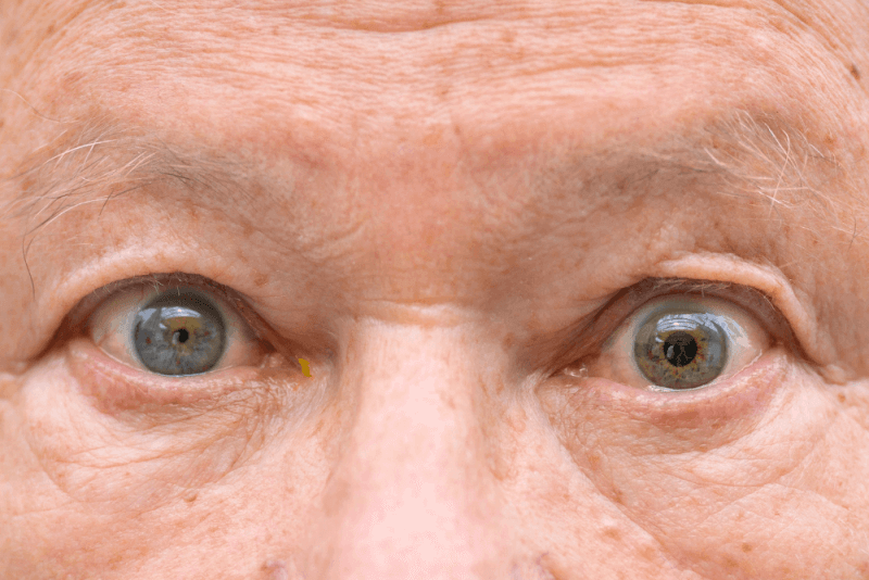
People with Sturge-Weber syndrome develop glaucoma, or eye pressure, in infancy or early adulthood. Glaucoma is a condition caused by increased eye pressure and leads to visual impairment.
In infancy, eye symptoms can sometimes be very pronounced. The pupils may appear large and swollen, and hemangiomas may be seen in different parts of the eyes. A hemangioma occurs when the blood vessels in the eye become enlarged and can cause vision loss in the future. In eye examinations, eye findings can be examined by measuring eye pressure and by examination.


C** C** | 21 Feb 2025