What is Ultrasound?
Ultrasound, also known as sonography or ultrasonography, is a non-invasive imaging test. The image produced by ultrasound is called a sonogram. Ultrasound uses high-frequency sound waves to create real-time images or videos of internal organs or other soft tissues like blood vessels.
With ultrasound, specialists can see the details of soft tissues inside the body without making any incisions. Unlike X-rays, ultrasound does not use radiation. Although often associated with pregnancy, specialists use ultrasound in many different situations to examine various internal parts of the body.
How does Ultrasound work?
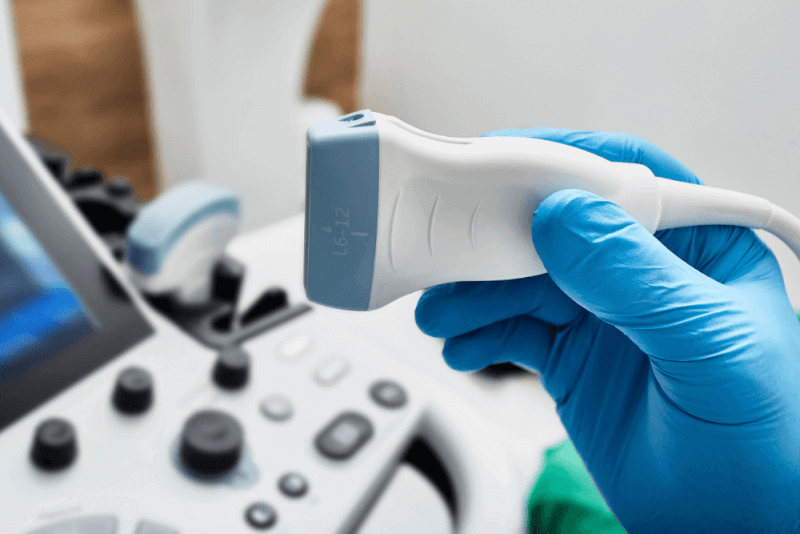
During an ultrasound, specialists move a device called a transducer or probe over an area of the body or inside a body cavity. They apply a thin layer of gel to the skin to ensure the ultrasound waves are transmitted from the transducer through the gel into the body.
The probe converts electrical currents into high-frequency sound waves and sends these waves into the body's tissues. The sound waves are inaudible. They bounce back from internal structures and return to the probe, which then converts them into electrical signals. A computer processes these signals into real-time images or videos displayed on a nearby computer screen.
Types of Ultrasound
There are various types of ultrasound imaging due to technological differences designed to address different issues more clearly. These variations in ultrasound devices have resulted in several types of ultrasound. The types of ultrasound include:
Color Doppler Ultrasound
Also known as color Doppler ultrasound, this advanced device is used to monitor blood flow. Color ultrasound imaging tracks blood flow in the vessels and investigates the causes of any narrowing in the vessels.
Widely used during pregnancy, color imaging provides safe visualization for areas like the legs, arms, brain, and liver. The uses of color Doppler include monitoring blood flow in the legs, arms, abdomen, or anywhere in the body, detecting heart valve diseases, vascular obstructions, vascular narrowing problems, blood flow issues in vessels, and during pregnancy.
- Legs, arms, abdomen, or anywhere in the body
- Heart valve diseases
- Vascular obstructions
- Vascular narrowing problems
- Blood flow issues in vessels
- Pregnancy
Detailed Ultrasound
Detailed ultrasound provides more detailed images to examine specific organs or systems within the body. It allows for detailed imaging of organ structures. It is often used for assessing abdominal or pelvic organs and monitoring pregnancy.
Doppler Ultrasound
Doppler ultrasound is used for diagnostic purposes. It evaluates the movement of substances like blood within the body. This allows doctors to see and assess blood flow in arteries and veins. Doppler ultrasound is often part of a diagnostic ultrasound study.
3D Ultrasound
3D ultrasound is typically used in pregnancy to obtain a three-dimensional image of the baby. It allows the visualization of specific birth defects, such as a cleft palate, that may not be seen with standard ultrasound.
4D Ultrasound
4D ultrasound is another type used during pregnancy. It provides a video of the 3D images, allowing the observation of real-time movements like yawning or stretching.
Elastography
Elastography is an imaging system that checks if organs are harder than normal. Hard areas in organs can indicate disease. Elastography is primarily used to check the stiffness of the liver, as hard areas in the liver can indicate scar tissue from liver disease.
Endoscopic Ultrasound
Endoscopic ultrasound is a minimally invasive procedure for evaluating diseases of the digestive system and other nearby organs and tissues. It combines the use of a thin, flexible tube placed into the gastrointestinal system with a device that uses sound waves to create images. High-frequency sound waves produce detailed images of surrounding organs and tissues, including the digestive system, lungs, pancreas, gallbladder, liver, and lymph nodes.
How is an Ultrasound performed?
Generally, no special preparation is required for an ultrasound test. However, for pelvic and urinary tract ultrasounds, including pregnancy ultrasounds, the bladder may need to be filled by drinking water before the test. For abdominal ultrasound, dietary adjustments or fasting may be required a few hours before the test.
Preparation for an ultrasound depends on the body part being scanned. Specialists may ask you to remove certain clothing or wear a hospital gown. The following general steps are involved for ultrasounds that involve applying the probe to the skin.
- Lie on your side or back on a comfortable table.
- The ultrasound technician applies a small amount of water-soluble gel to the skin over the area to be examined. This gel does not harm the skin or stain clothes.
- The technician moves a hand-held transducer or probe over the gel to obtain images inside the body.
- During the imaging, the technician may ask the patient to remain still or hold their breath for a few seconds to get clearer images.
- After enough images are obtained, the remaining gel is wiped off, and the test is completed.
When is a detailed ultrasound performed?
Ultrasound imaging is often used during pregnancy to closely monitor the health of the fetus and the mother. Different types of ultrasound scans are performed at various stages to diagnose different issues early.
One such ultrasound is the color Doppler ultrasound, which is often requested by specialists to check the development of blood vessels. The best times to get clear images are between the 11th and 13th weeks and between the 18th and 21st weeks of pregnancy.
Another ultrasound method used to closely monitor pregnancy is the detailed ultrasound. It is performed between the 18th and 22nd weeks of pregnancy and is commonly referred to as the 20-week ultrasound or pregnancy ultrasound.
What conditions can ultrasound detect?
Ultrasound is frequently used to diagnose a variety of medical issues, including:
- Abnormal growths such as tumors or cancer
- Blood clots
- Enlarged spleen
- Ectopic pregnancy
- Gallstones
- Aortic aneurysm
- Kidney or bladder stones
- Cholecystitis
- Enlarged veins in the testicles
What are the uses of Ultrasound?
Ultrasound imaging is primarily used in three main categories. These categories include:
- Pregnancy ultrasound
- Diagnostic ultrasound
- Guidance for procedures
Pregnancy ultrasound
Doctors often use ultrasound to monitor the mother and fetus during pregnancy. The purposes of prenatal ultrasound include:
- Confirming the pregnancy
- Checking for multiple fetuses
- Estimating the gestational age
- Monitoring fetal growth and position
- Viewing fetal movement and heartbeat
- Checking for congenital conditions in the fetal brain, spine, heart, or other parts of the body
- Checking the amount of amniotic fluid
Most specialists recommend an ultrasound at 20 weeks of pregnancy. This test monitors the growth and development of the fetus during pregnancy. It also helps determine the fetus's biological sex. Additionally, specialists may request extra scans in response to any concerns or potential congenital conditions.
Diagnostic ultrasound
Doctors use diagnostic ultrasounds to view the internal parts of the body to see if something is wrong or not working correctly. With ultrasound, specialists can learn the cause of unexplained pain, lumps, and abnormal blood tests.
Most diagnostic ultrasound exams involve the technician placing the probe on the skin. In some cases, the probe may need to be inserted into the body, such as in the vagina or rectum. The specific diagnosis, type of ultrasound, and details of the condition determine this. Examples of diagnostic ultrasound include:
Thyroid ultrasound
Doctors use ultrasound to evaluate the thyroid, a butterfly-shaped endocrine gland in the neck. Ultrasound imaging can measure the thyroid and detect nodules or lesions within the gland.
Transrectal ultrasound
Specialists insert an ultrasound probe into the rectum. It is used to evaluate the rectum or prostate and nearby tissues in individuals assigned male at birth.
Breast ultrasound
Breast ultrasound is a non-invasive test for detecting cysts in the breast. It is often requested after an abnormal mammogram.
Hip ultrasound for babies
Hip ultrasound is performed to investigate dislocated or underdeveloped hips in infants. It is used from the newborn period until about 6-8 months of age. This imaging shows whether the baby's hip socket is well-formed and properly positioned.
Abdominal ultrasound
An ultrasound probe is moved over the skin in the belly area. Abdominal ultrasound can diagnose many causes of abdominal pain.
Kidney ultrasound
Also known as renal ultrasound, this test allows specialists to evaluate the size, location, and shape of the kidneys, ureters, and bladder. Ultrasound detects cysts, tumors, obstructions, or infections within or around the kidneys.
Pelvic ultrasound
Pelvic ultrasound evaluates the organs in the pelvic area between the lower abdomen and the thighs. Some pelvic organs include the bladder, prostate, rectum, ovaries, uterus, and vagina.
Transvaginal ultrasound
Specialists insert a probe into the vaginal canal to examine reproductive tissues like the uterus or ovaries.
Stomach ultrasound
Also known as abdominal ultrasound, it is used to view the inside of the abdomen. Abdominal ultrasound is preferred for diagnosing or ruling out problems if any issues arise.
Heart ultrasound
Known as an echocardiogram, this ultrasound creates images of the heart. It shows blood flow through the heart valves and is used to diagnose heart disease. Different types of heart ultrasounds are available, and the appropriate type is used depending on the problem. Some heart ultrasounds are performed during exercise or pregnancy.
Ultrasound guidance for procedures
Specialists sometimes use ultrasound to accurately perform certain procedures. A common use of ultrasound is to guide needle placement for taking fluid or tissue samples. Areas where tissue samples can be taken with ultrasound guidance include:
- Tendons
- Joints
- Muscles
- Cysts or fluid collections
- Soft tissue masses
- Organs
Procedures that may require ultrasound guidance include:
- Embryo transfer during IVF treatment
- Nerve blocks
- Confirmation of IUD placement after insertion
- Lesion localization procedures