What is an Arteriovenous Malformation (AVM)?
Also known as an arteriovenous malformation, an AVM is a blood vessel disorder that irregularly connects arteries and veins, disrupting blood flow and oxygen circulation. The function of arteries is to carry blood from the heart to the body and brain. Veins return oxygen-depleted blood cells from the tissues back to the heart.
In AVM patients, this flow is disrupted. As a result, sufficient oxygen cannot be delivered to the tissues. Additionally, due to the knot, the blood vessels may weaken and rupture. If an AVM occurs in the brain and the vessels rupture, it can lead to a brain hemorrhage, stroke, and brain damage.
Arteriovenous Malformation (AVM) Diagnostic Criteria
In the diagnosis of AVM, a physical examination is initially performed. During the physical examination, the presence of a sound called a bruit is investigated. A bruit is a whistle-like sound caused by the very rapid blood flow in the vessels and arteries due to an AVM.
This sound can also be heard by patients, potentially affecting their sleep patterns. Other tests used in the diagnosis of AVM include the following:
- Cerebral angiography allows the structure of the vessels to be seen more clearly under X-rays.
- CT scan
- CT angiography can detect areas of bleeding.
- MRI
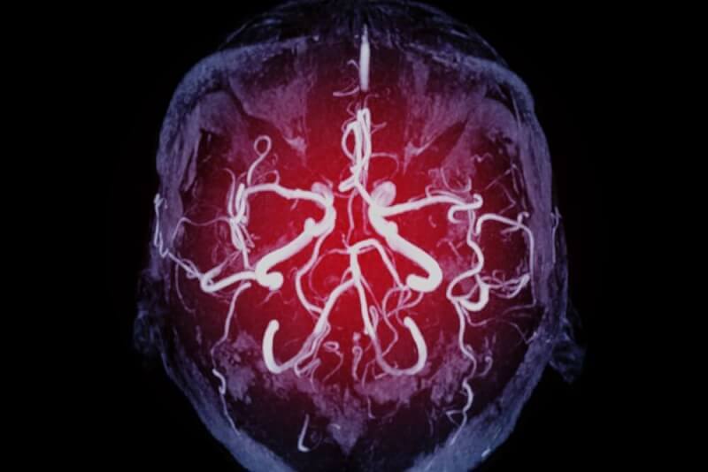
- MRA can detect the shape, speed, and distance of blood flow in irregular vessels.
Arteriovenous Malformation (AVM) Symptoms
The symptoms seen in a patient vary depending on the location of the AVM. In a significant portion of cases, symptoms appear after bleeding occurs. In addition to bleeding, other symptoms that may occur include:
- Loss of consciousness
- Progressive loss of neurological function
- Seizures
- Headache
- Nausea
- Vomiting
Other symptoms that may occur with an AVM include:
- Weak muscles
- Paralysis in one part of the body
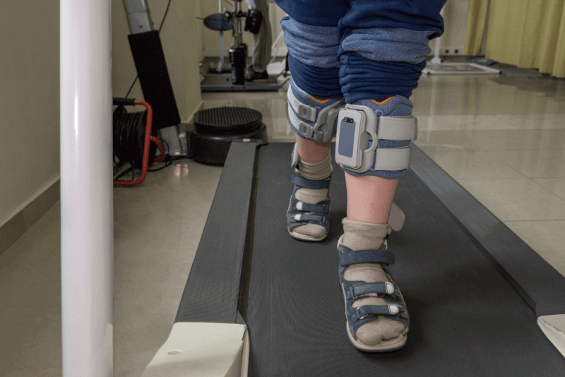
- Coordination loss that may cause difficulty walking
- Difficulty performing tasks that require planning
- Weakness in the lower extremities
- Back pain
- Dizziness
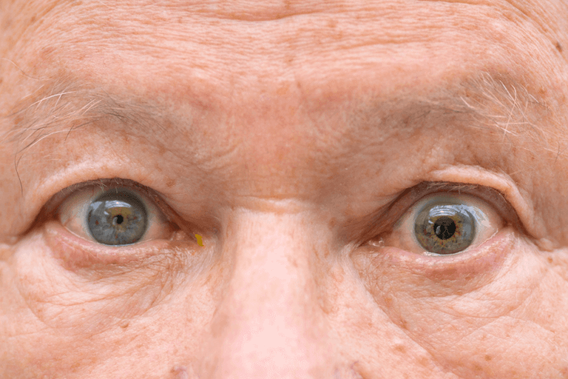
- Partial loss of vision
- Loss of control over eye movements
- Swelling of part of the optic nerve
- Speech and comprehension disorders
- Numbness, tingling, and sudden pain
- Memory loss
- Dementia
- Hallucinations
- Confusion
- Disorientation
In children, an AVM may cause learning and behavioral disorders.
A type of AVM called a Galen vein malformation presents with symptoms at birth or shortly thereafter. In the Galen defect, the AVM forms deep within the brain. Symptoms of a Galen vein malformation may include:
- Fluid buildup in the brain causing an enlarged skull
- Swollen veins on the scalp
- Seizures
- Developmental delays
- Congestive heart failure
Causes of Arteriovenous Malformation (AVM)
AVM is caused by the development of irregular connections between arteries and veins. However, the underlying reasons for this change are unknown. Some genetic changes are thought to play a role in the occurrence of AVM, although not all cases present with these genetic factors.
Arteriovenous Malformation (AVM) Complications
Complications that may occur with AVM include:
Brain Hemorrhage
The most significant risk of AVM is brain hemorrhage. Bleeding resulting from AVM can lead to stroke, brain damage, or seizures.
Seizures
Disruptions in electrical activity in the brain can cause fainting and uncontrolled muscle movements.
Aneurysm
Weakening of the blood vessels due to AVM can also lead to an aneurysm. Approximately 50% of cases experience an aneurysm, increasing the risk of bleeding and the severity of symptoms.
Brain Damage
It can affect mental processing, thinking, memory, speech, and comprehension.
Other Complications
In addition to the above complications, coma and death may occur in patients with AVM.
Treatment Methods for Arteriovenous Malformation (AVM)
The treatment of AVM is planned based on the location of the issue and the symptoms. Under normal circumstances, changes in AVM patients are monitored with regular imaging tests. If treatment for AVM is necessary, the following signs are observed:
- Bleeding
- Non-bleeding symptoms
- The treatability of the area where the AVM is located
Arteriovenous Malformation (AVM) Surgery
The primary treatment for AVM is surgical procedures. Surgery is the only method that ensures complete treatment of the condition. However, these methods are applied in situations where the procedure can be performed without damaging the brain tissue.
Surgical Methods for Arteriovenous Malformation (AVM)
Surgeries for AVM are called endovascular embolization. In this procedure, surgeons pass a catheter through the arteries to the AVM area. Then, a special drug is injected into the part of the AVM where the catheter remains, reducing blood flow. This method also reduces the risk of complications.
In some cases, stereotactic radiosurgery is used to treat AVM. These procedures involve using focused radiation beams to damage the vessels and stop the blood flow in the knot.
Side Effects of Arteriovenous Malformation (AVM) Surgery
Possible side effects after AVM surgery may include:
- Bleeding
- Headache
- Swelling
- Damage to nearby tissues
- Muscle weakness on one side
- Effects on speech, hearing, and vision
- Disabling or life-threatening complications
Life After Arteriovenous Malformation (AVM) Surgery
After surgery, it must be confirmed that there is no bleeding in the AVM area. For this purpose, an angiogram is performed. Patients are then transferred to the intensive care unit. After the surgery, patients are monitored for several days.
If necessary during this period, patients may need to start a rehabilitation process to regain their strength, speech, and other functions. After the surgery, patients may experience pain in the scalp. Additionally, numbness around the incision, swelling, and bruising around the eyes may also occur.
The recovery process lasts between 4 to 6 weeks, during which patients may feel more tired than usual. After 6 weeks, patients can start returning to normal activities, but it may take between 2 to 6 months to fully return to their normal life.
What Should Individuals with Arteriovenous Malformation (AVM) Pay Attention To?
Even after treatment, AVM can recur. Therefore, if symptoms reappear after treatment, it is important to consult a doctor. Additionally, patients should pay attention to the following points:
- After surgery, patients should attend follow-up appointments every three months.
- After the first year, annual follow-up appointments are sufficient.
- In addition, if symptoms such as seizures, sudden and severe headaches, weakness in the arms or legs, vision problems, balance issues, memory problems, or attention difficulties occur, it is necessary to visit the emergency room.
What Are the Risk Factors for Arteriovenous Malformation (AVM)?
Arteriovenous malformations are rarely inherited. However, having a family history of AVM increases the risk. Additionally, some hereditary conditions such as Osler-Weber-Rendu syndrome increase the risk of developing an AVM.
Where Do Arteriovenous Malformations (AVM) Occur?
Arteriovenous malformations can occur anywhere in the body, and most do not cause any health problems. However, AVMs in the brain and spinal cord can cause serious health issues and symptoms.
Liver Arteriovenous Malformation
Liver hemangiomas are benign masses formed from blood vessels in the liver. They are a very common condition and are thought to affect approximately 20% of the population.
Liver AVMs rarely cause symptoms and are usually diagnosed through imaging studies performed for other issues. There is no evidence that liver AVMs cause cancer, and treatment is usually not required.
Liver Arteriovenous Malformation Symptoms
If liver AVM symptoms occur, they may include:
- Pain in the upper right abdomen
- Feeling full after eating a small amount
- Nausea
- Vomiting
Brain Arteriovenous Malformation
Brain AVMs are among the most dangerous types of AVMs. Although these masses are not cancerous, they can cause many different symptoms. Often, no symptoms are seen, but serious symptoms can occur if bleeding or rupture occurs.
Skin Arteriovenous Malformation
The most common location for AVMs is the skin. Skin AVMs appear as bright red nodules and are often considered birthmarks. They are especially common on the face, scalp, back, and chest.
Skin AVMs, also known as vascular birthmarks, grow during a baby’s first year. They then enter a resting phase and usually heal by age 5. By the time children reach age 10, they are barely noticeable.


