30 Second Summary
- Gliomas are tumors that form in glial cells, the supporting tissue of the brain.
- It accounts for about 30% of all brain tumors.
- It is classified as benign (low grade) and malignant (high grade).
- Symptoms vary depending on the size, location and grade of the tumor.
- Treatment includes surgery, radiation therapy and chemotherapy.
What is a glioma?
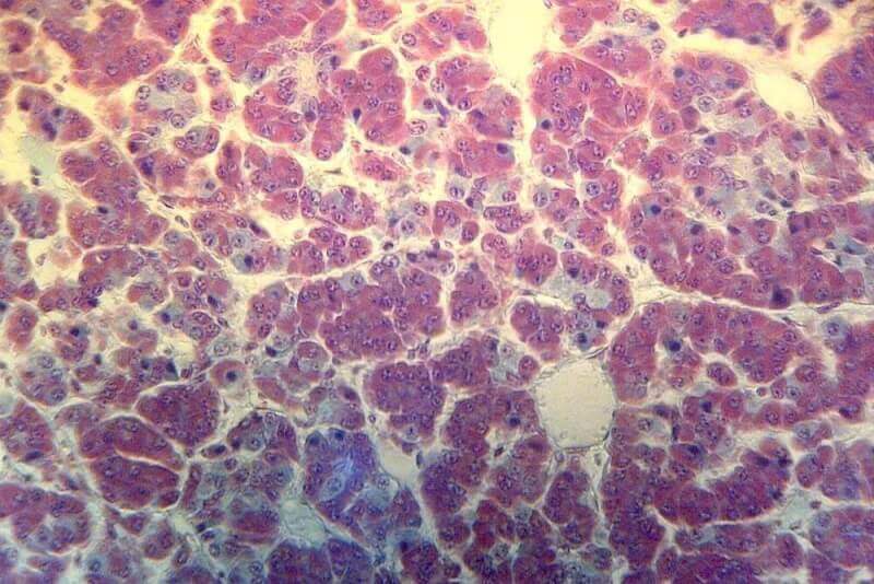
Glioma is a tumor that can occur in the brain and spinal cord of both adults and children. It is caused by the controlling protrusion of glial cells. Glial cells support the nerves, helping the central nervous system to function under normal conditions. Gliomas usually occur in the brain. However, there are cases where it is also seen in the vertebral marrow.
Some types of gliomas, which are malignant tumors, grow very slowly. Since they originate from brain tissue, they are primary brain tumors. Gliomas that do not usually spread outside the brain and spine can be life-threatening. Among the reasons why glioma is so effective; it is seen in hard-to-reach and hard-to-surgery areas and it grows towards other parts of the brain.
Glioma diagnostic criteria
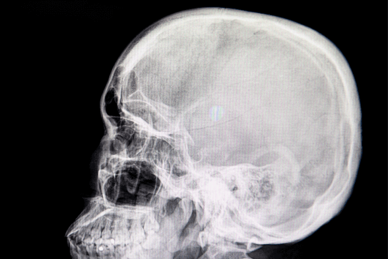
To diagnose glioma, specialists first review the patient's symptoms and medical history. A complete physical and neurological examination is then required. MRI and CT scans are particularly useful in glioma diagnosis. If any abnormal mass is detected during these scans, a biopsy is performed. Among the reasons why a biopsy is performed are the following.
- Whether the mass is cancerous
- The tumor is not the result of an abnormal gene
- Type of cell in the tumor
- How aggressive the tumor is
Causes of glioma
Studies on glioma show that changes or mutations in DNA cause cells to proliferate out of control. These genetic mutations can be inherited from parents or they can appear suddenly during the course of life.
Glioma symptoms
Symptoms of glioma include the following.
- Speech and communication problems
- Changes in vision
- Difficulty in learning, thinking and remembering
- Difficulty walking
- Difficulty maintaining balance
- Dizziness
- Headache
- Weakness and numbness on one side of the body
- Nausea
- Vomiting
- Personality changes
- Behavior changes
- Seizures
Glioma treatment methods
Glioma treatment methods include multiple options. Factors influencing the planning of treatment include the following.
- General health status of the patient
- Age of the patient
- Location of the tumor
- Type of tumor
- Size of the tumor
- Whether there has been treatment for brain cancer in the past
The main method of glioma treatment is surgical methods. Tumors, especially those that are easily accessible to the surgeon, are surgically removed. However, for tumors that are difficult to remove or close to sensitive areas, additional treatments such as radiotherapy and chemotherapy are necessary.
Chemotherapy treatment
Chemotherapy drugs used to destroy cancer cells are used in the treatment of glioma as well as many other types of cancer. Temozolomide is the most commonly used drug in oral or intravenous chemotherapy sessions. This medicine also increases the effectiveness of radiation therapy.
Radiotherapy treatment
Radiation therapy used in the treatment of glioma is applied using the strongest dose of radiation to destroy tumors. Radiation therapy, which is used especially in hard-to-reach places, can be determined as the primary treatment method or it can be used to shrink the tumor. In radiation therapy, the exact shape of the tumor is targeted and the risk of damage to surrounding tissues is minimized.
A type of radiation therapy called brachytherapy can also be applied. In this treatment, radiation sources are placed near the tumor. These sources then emit radiation without harming nearby healthy tissues.
Glioma surgery
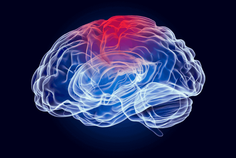
During surgery, surgeons use special techniques such as imaging and brain mapping to guide the operation. Brain mapping shows which areas of the brain control which vital functions. With this information, the surgeon can remove the tumor without damaging healthy tissue.
Glioma treatment usually starts with surgery. If the entire tumor is removed with these surgeries, it remains the only surgical procedure. However, in some cases it is not possible to remove the entire tumor. The surgeon removes as much tumor as possible. This procedure is also called total resection. Total resection is needed especially if the tumor cannot be easily separated from the brain tissue. The reason for performing a total resection is that it causes a significant reduction in the patient's symptoms.
Methods of glioma surgery
Glioma surgeries can be performed with different techniques depending on the size and location of the tumor. Open brain surgery called craniotomy is the most common type of surgery used to remove gliomas.
If the size and location of the tumor allows, it is possible to remove the tumor with the ablation technique. Ablation is a minimally invasive surgical technique that uses heat from a laser to destroy part or all of the brain tumor.
Endoscopic head surgery
It is one of the minimally invasive brain surgeries that is gaining popularity in brain surgery. To remove a glioma in the frontal part of the brain, access is made through the nose into the skull. However, in order to perform this surgery successfully, the surgeon must be highly experienced in the field.
Intraoperative imaging and surgical navigation
Intraoperative imaging and surgical navigation are the latest and most technologically advanced methods used in glioma surgery in centers specializing in neurosurgery. This method allows neurosurgeons to guide their interventions in real time as they remove the tumor. This system, which provides better visualization of tumor spread with brain ultrasonography and MRI, also ensures more successful surgeries.
Intraoperative neuromonitoring
It is one of the latest technologies applied in neurosurgery. With this method, the surgical team uses various devices to measure electrical activity in different parts of the brain. With these devices, nerve pathways such as hearing and vision can be identified. This allows the surgeon to go around these nerve pathways during the operation to minimize damage to the nerves. In order for the surgeries to take place, the support of specialized neurophysiologists is needed in the operating room. Intraoperative neuro-monitoring procedures can be performed in different types. These include the following.
- Bodily sensory evoked potentials used to identify areas responsible for deep and surface emotions
- Acoustically evoked potentials or auditory evoked potentials for testing hearing
- Visual evoked potentials for the identification of visual neural pathways
- Electromyography for identification of important cranial nerves
- Electroencephalography for the assessment of spontaneous electrical activity in brain cells
- Direct cortical stimulation to identify the neural pathways responsible for controlling movement
- Direct nerve stimulation
Complications of glioma surgery
Glioma surgeries carry some risks. The most important risks are infection and bleeding. Other complications that may occur after surgery vary depending on the location of the tumor. For example, during the removal of a tumor located close to the optic nerves, the optic nerves may be damaged, causing the patient to lose vision.
Treatment of tumor treatment areas
Tumor treatment fields therapy uses electrical energy to damage glioma cells. This treatment makes it harder for new glaucoma cells to be made. It is especially applied to patients with the highly aggressive glioblastoma type of glioma.
Pads are used in therapies to treat tumor areas. The pads are glued to the scalp with hair. It may be necessary to shave the hair so that the pads can be made precisely. The wires coming out of the device are then connected to the pads. The energy of the device generates an electric current that damages the glioma cells. The side effect of this treatment is that it can cause skin irritation in the areas where the pads are applied to the head terrace.
Targeted therapy
Targeted therapy, which focuses on specific chemicals found in cancer cells, ensures that cancer cells die. Glioma cells need to be tested to determine whether targeted therapy will be beneficial for the patient. It is a treatment option after surgical methods, especially for gliomas that develop slowly and cannot be completely removed. Side effects in targeted therapy vary depending on the drug and dose.
The healing process of glioma
The recovery process after glioma surgery differs for each patient. While some patients can resume their daily routine a few weeks after surgery, some patients require a more comprehensive rehabilitation plan. Successful recovery after glioma removal surgery requires a comprehensive recovery plan and multidisciplinary cooperation. For this, the following disciplines working together will ensure that the patient will recover in a short time.
- Physical therapy
- Occupational therapy
- Speech therapy
- Pain management
- Counseling for patients and caregivers
Risk factors of glioma disease
Glioma is one of the diseases that can be seen in everyone. However, the following factors increase the risk of occurrence.
- Over 65 years of age
- Under 12 years of age
- Belonging to the white race
- Some genetic disorders in the family
- Becoming a man
Types of glioma
It is divided into 3 main types according to the type of glial cell it starts from. In some cases, more than one cell type is seen in tumors. These conditions are called mixed gliomas. Each type of glioma is classified as low, intermediate or high grade according to its growth rate and other characteristics.
Astrocytomas
Astrocytomas that start in cells called astrocytes include glioblastomas and diffuse intrinsic pontine gliomas. Glioblastomas are one of the fastest growing astrocytoma types because they are extremely aggressive. The most common malignant brain tumor in adult patients is astrocytoma. In children, although it is a rare form of brain cancer, it is extremely aggressive and usually occurs in the brain stem.
Oligodendrogliomas
Oligodendrogliomas, which start in glial cells called oligodendrocytes, tend to grow more slowly than other types. They can also become aggressive in the later stages of the disease. Like ependymomas, they rarely spread outside the brain and spine. It is more common in adults than in children. The incidence of oligodendroglioma, including all other brain tumors, is between 1% and 2%.
Ependymomas
Ependymomas, which are formed in ependymocytes, one of the glial cell types, usually occur in the ventricles of the brain and spinal cord. They can spread through the fluid that surrounds and protects the brain and spinal cord. However, they are not seen outside the brain and spine. Ependymomas, which account for approximately 2% of all brain tumors, are more common in children than in adults.
Glioma complications Complications
Life-threatening complications of gliomas include
- Brain hemorrhage
- Brain hernia, which is defined as the brain tissue moving out of its normal position in the skull
- Accumulation of fluid in the brain, called hydrocephalus
- Increased pressure inside the skull
- Seizures
How to prevent glioma?
A significant proportion of the risk factors that cause glioma cannot be controlled. However, by detecting gliomas at an early stage, the progression of the disease can be slowed down and treatment can be successful. Genetic testing is especially recommended for people with a family history of brain tumors. It would be useful to discuss the risks and benefits of genetic testing with your physician. In addition, avoiding radiation exposure to the head and leading a healthy lifestyle will help prevent glioma.