30-Second Summary
- A significant portion of meningiomas are benign. However, they can sometimes be malignant. If a tumor is cancerous or malignant, there is a possibility that other tissues may also be invaded.
- Meningiomas are usually located near the upper and outer curve of the brain. They can also form at the base of the skull. Spinal meningiomas are rarely seen.
- The exact cause of meningiomas is not yet known. Studies have shown that 40% to 80% of all meningioma cases have an abnormality in chromosome 22, which plays a role in suppressing tumor growth.
- It may not always be possible to diagnose a meningioma. Due to the extremely slow growth of the meningioma, symptoms may not appear immediately. Usually, symptoms arise after the tumor has grown significantly and started to press on nearby tissues.
What is a Meningioma?
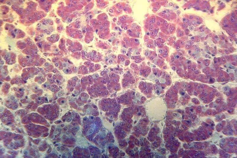
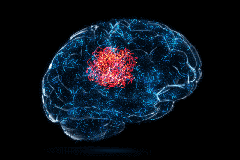
A meningioma, one of the types of tumors, occurs in the meninges. The meninges are tissues that cover the brain and spinal cord and help protect these structures. The most common cell type in which meningiomas occur is arachnoid cells. These cells are found within the thin, spider web-like membrane that covers the brain and spinal cord. It is one of the three layers that make up the meninges.
A significant portion of meningiomas are benign. However, they can sometimes be malignant. If a tumor is cancerous or malignant, there is a possibility that other tissues may also be invaded. Therefore, a meningioma may potentially spread to other tissues in the body. However, if the tumor is benign, there is no possibility of it spreading to other tissues in the body.
Meningiomas are usually located near the upper and outer curve of the brain. They can also form at the base of the skull. Spinal meningiomas are rarely seen.
The characteristic feature of benign meningiomas is their tendency to grow slowly. In addition, the growth occurs inward. Therefore, benign meningiomas can reach extremely large sizes before being diagnosed. Even benign meningiomas can grow large enough to threaten life if they become compressed near areas of the brain and exert pressure on them.
Meningiomas, one of the most common types of tumors seen in the head, are not brain tumors. However, they can exert pressure on nearby brain tissue, nerves, and blood vessels. A significant portion of meningiomas grow very slowly. Therefore, they can grow for years without causing symptoms. Meningiomas are more commonly seen in women. Meningiomas tend to occur more frequently in women, especially in older women.
Causes of Meningioma
The exact cause of meningiomas is not yet known. Studies have shown that 40% to 80% of all meningioma cases have an abnormality in chromosome 22, which plays a role in suppressing tumor growth. Genetic conditions are rarely seen in meningioma cases.
In addition, it is generally a health issue that arises spontaneously. Environmental, genetic, and hormonal risk factors are known to be among the causes of meningioma.
Symptoms of Meningioma
Since meningiomas are generally slow-growing tumors, they may not cause noticeable symptoms until they grow large enough to press against important surrounding structures. The symptoms of meningiomas vary greatly depending on which part of the brain is affected.
Certain meningioma locations are associated with specific neurological symptoms. Examples include the following.
- Olfactory meningiomas may lead to partial or complete loss of the sense of smell.
- Tumors located on the posterior frontal midline may cause paralysis of the upper parts of the legs or the lower part of the body.
- Sphenoid wing meningiomas may cause one or both eyes to be positioned outside of their normal alignment.
The most common symptoms of meningiomas include the following:
- Paralysis in certain parts of the body
- Headache
- Muscle weakness in specific areas
- Dizziness
- Overactive reflexes
- Nausea
- Hyperreactive reflexes
- Vomiting
- Memory problems
- Double vision
- Behavioral changes
- Hearing loss
- Personality issues
- Seizures
- Vision loss
The most common symptoms of spinal meningioma include the following:
- Pain at the location of the tumor
- Radiculopathy
- Weakness
- Decreased muscle tone
- Neurological problems such as reduced or lost reflex responses
Meningioma Diagnostic Criteria
It may not always be possible to diagnose a meningioma. Due to the extremely slow growth of the meningioma, symptoms may not appear immediately. Usually, symptoms emerge after the tumor has grown significantly and begins to press on adjacent tissues. Additionally, since most meningiomas grow slowly, they tend to affect adults.
In some cases, symptoms may be so mild that patients might think they are a normal part of life. If a meningioma is suspected, patients are referred to a neurologist. A physical and neurological examination is performed to diagnose a meningioma. In addition, the following imaging tests are also recommended.
Brain Magnetic Resonance Imaging (MRI)
The best imaging test to diagnose a meningioma is a contrast-enhanced brain MRI scan. Contrast-enhanced MRI helps improve the diagnostic quality of the images. This test is performed using a large magnet and is part of imaging tests. The radio waves used also allow visualization of tissues and organs inside the body.
Computed Tomography (CT) Scan
For those who cannot undergo an MRI, specialists will likely recommend a contrast-enhanced head CT scan. Computed tomography scans use X-rays. Combined with computer technology, it allows detailed imaging of body structures. Sometimes, a contrast agent—often called dye—helps enhance the images by highlighting certain features. The contrast agent can be taken orally or injected intravenously.
In some cases, imaging tests alone may not be sufficient. In such situations, a biopsy may be performed to confirm the diagnosis. A biopsy also helps determine whether the tumor is cancerous.
Treatment Methods for Meningioma
Treatment for meningioma is highly personalized and will most likely include a combination of the following treatments.
External Beam Radiation Therapy
External beam therapies are also referred to as EBRT and are the most commonly used method among these types of treatments. In this therapy, high-energy radiation beams are directed at the target to help protect more of the healthy tissue.
Brachytherapy
Another radiation option used in cancer treatment is brachytherapy. In this method, radiation is applied directly inside the tumor. Radioactive capsules, seeds, or other implants are used for this purpose. In some cases, it may not be possible to apply the radioactive substance directly into the tumor. In such situations, it is placed in the location closest to the tumor.
Fractionated Stereotactic Radiotherapy (SRT)
This type involves delivering small fractions of radiation over time, such as one treatment per day for 30 days. With this approach, it can be used for tumors that are too large for surgery or located in areas where radiosurgery is particularly effective, such as tumors near the optic nerves.
Intensity-Modulated Radiation Therapy (IMRT)
This treatment uses computer software to reduce the intensity of radiation delivered to the meningioma area. It is used for meningiomas that are close to sensitive brain structures or have a complex shape.
Gamma Knife Radiosurgery
This treatment takes place in an outpatient setting. Therefore, hospital stay is not required. Many patients need only one treatment session. However, depending on the size and location of the treated area, it may take up to 5 sessions. During the procedure, approximately 200 low-dose radiation beams are used to directly treat the meningiomas.
Since the Gamma Knife system is highly precise, the doctor can treat an area as small as 0.15 mm and protect surrounding healthy tissues from the effects of radiation. This means fewer of the unpleasant side effects typically associated with radiation therapy.
For atypical and cancerous meningiomas, adjuvant radiotherapy improves long-term tumor growth control, survival, and overall survival. Sometimes referred to as supportive therapy, adjuvant therapy targets cancer cells that the primary treatment could not eliminate. This therapy helps destroy cancer cells that are not affected by the primary treatment method. After complete surgical removal of atypical meningiomas, adjuvant radiotherapy reduces the risk of tumor recurrence.
Side Effects of Radiation Therapy
Although the radiation treatment process for meningioma is not painful by itself, it may cause some side effects due to healthy tissues being exposed to radiation. The most common side effects of radiation therapy for meningioma include the following:
- Mild skin reactions
- Hair loss
- Fatigue
- Difficulty in thinking clearly
- Mild memory loss
- Loss of appetite
- Headaches
A significant portion of the side effects caused by radiotherapy are temporary and therefore disappear after the end of treatment.
Meningioma Surgery
When planning meningioma surgery, neurological examination, MRI, and CT scans must be reviewed. Depending on the location of the tumor, it may involve major blood vessels. In such cases, an angiogram may be performed to observe how the tumor affects these vessels and to identify the vessels supplying blood to the tumor.
An angiogram is a type of X-ray that produces images of the blood vessels supplying oxygen and other nutrients to the meningioma and helping it grow. Before the test, a contrast dye is injected through a catheter into an artery going to the brain to produce more detailed images of the tumor and surrounding blood vessels.
In some cases, doctors may inject a substance that blocks blood flow to the tumor. This procedure, called embolization, is performed during the angiogram to reduce the amount of bleeding during surgery. These procedures are performed by surgeons known as interventional neuroradiologists. For spinal meningioma, angiography is rarely needed because these types of tumors generally do not involve critical blood vessels.
During meningioma surgeries, image guidance is used. Image guidance is used to precisely locate a tumor beneath the skull. It ensures a sufficiently large and precisely positioned opening directly above the tumor is created in the skull, preventing unnecessary exposure of other brain areas.
Image guidance is provided by a computer using MRI and CT scans during surgery. This approach can also be used to remove meningiomas from the spinal cord. Neurosurgeons also use ultrasound to locate spinal cord tumors and guide the procedure.
During surgeries involving the brain and spinal cord, surgeons receive real-time information about the patient's neurological function, which helps minimize damage and injury to nearby nerves, the brain, and the spinal cord. Doctors who provide this special monitoring are called neurophysiologists. They remain in the operating room throughout the surgery.
Methods of Meningioma Surgery
The surgery to be performed varies depending on the size and location of the tumor. The following are among the meningioma surgery methods.
Craniotomy
For a meningioma located on the surface of the brain, surgeons may perform a procedure called a craniotomy, which involves opening the skull. Craniotomy can also be used for tumors located near the base of the skull, such as those close to the brainstem where the brain meets the spinal cord.
Craniotomy is performed under general anesthesia. The surgeon makes an incision in the scalp and removes a small part of the skull to create an opening. A surgical microscope is typically used to provide high magnification of the tumor and surrounding critical structures. Using micro-instruments—very small tools used for performing complex surgeries—the surgeon removes as much of the tumor as possible.
The surgeon then closes the hole with the removed portion of the skull and stitches the skin together.
Life After Meningioma Surgery
The hospital stay after meningioma surgery can vary from a few days to several weeks depending on the size of the tumor, its location, and the type of procedure used to remove it. For example, minimally invasive procedures such as endoscopic endonasal or neuroendoscopic surgery may result in shorter recovery times compared to craniotomy or spinal surgery.
During recovery, doctors ensure that patients are as comfortable as possible. They may prescribe various medications to help alleviate side effects such as nausea or headaches. Additionally, they monitor brain and spinal nerve functions to determine if any issues related to cognition or mobility arise as a result of the surgical intervention.
Palliative Care
Meningioma and its treatment can cause physical symptoms and side effects as well as emotional and social problems. The treatment aimed at managing these effects is called palliative care. Palliative care, used alongside treatments designed to slow, stop, or eliminate the tumor, is an important part of patient care.
Palliative care focuses on managing symptoms and supporting the patient and their family. While there are many different approaches to palliative care, it often includes the following:
- Nutritional changes
- Medications
- Emotional and spiritual support
- Relaxation techniques
- Procedures aimed at improving neurological function
- Procedures aimed at improving quality of life
Chemotherapy
Chemotherapy, one of the options used in cancer treatment, involves the use of various drugs to kill cancer cells. Although the use of chemotherapy in the treatment of meningiomas is rare, specialists often recommend it for individuals who develop recurrent or progressive meningiomas that no longer respond to surgery and radiation therapy.
In patients with anaplastic meningiomas, certain types of chemotherapy have shown successful results in tumor regression following surgical resection and radiotherapy.
The possible side effects of chemotherapy include the following:
- Neurofibromatosis type 2
- Von Hippel-Lindau disease
- Multiple endocrine neoplasia type 1
- Li-Fraumeni syndrome
- Cowden syndrome
Meningioma Complications
Meningioma or its treatment can lead to long-term complications. Treatment usually involves surgical intervention and radiation therapy. Possible complications include:
- Difficulty concentrating
- Memory loss
- Personality changes
- Seizures
- Weakness
- Sensory changes and speech difficulties
Meningioma Grades
Meningiomas are classified into three grades:
- Grade I (typical): A group of slow-growing, benign meningiomas. Typical meningiomas account for approximately 80% of cases.
- Grade II (atypical): These meningiomas grow more rapidly, are more resistant to treatment, and are not cancerous. These tumors affect approximately 17% of patients.
- Grade III (anaplastic): A fast-growing, malignant form that spreads quickly. These tumors account for about 1.7% of cases.
Additionally, meningiomas are classified into several types based on location and tissue type. Examples of locations include:
- Convexity meningiomas that grow on the surface of the brain and may apply pressure as they enlarge
- Intraventricular meningiomas that grow within the brain’s ventricles
- Olfactory groove meningiomas that occur at the base of the skull between the brain and nose, near the olfactory nerve
- Sphenoid wing meningiomas that form along the bony ridge behind the eyes
What Should Individuals With Meningioma Keep in Mind?
A meningioma diagnosis can be life-changing. The treatment process often involves frequent visits to doctors and surgeons. To help patients cope, the following strategies may be beneficial:
Learning About Meningiomas
It’s important to find reliable sources of information about meningiomas and treatment options. Consulting with experts can help identify good information sources, including local libraries or online resources. It is also recommended that patients write down any questions they have before visiting their doctor.
Building a Support Network
Having someone to talk to about their feelings is crucial. Other individuals who can provide support include social workers and psychologists. Doctors may recommend suitable professionals. Talking to spiritual leaders can also provide the support patients need.
Meeting or joining groups of people who are dealing with the same illness can offer emotional support and help patients feel less alone in their journey.
Taking Good Care of Themselves
Patients are advised to maintain a diet rich in fruits and vegetables. With a healthcare provider’s approval, engaging in moderate daily exercise can help improve overall health. Additionally, getting adequate, quality sleep should be a priority.
It is crucial for patients to reduce stress and focus on what is most important to them.
Meningioma Survival Rates
Studies on 5-year survival rates for meningiomas have found the following results:
- Grade I: 95.7%
- Grade II: 81.8%
- Grade III: 46.7%
10-year survival rates for meningiomas are as follows:
- Grade I: 90%
- Grade II: 69%