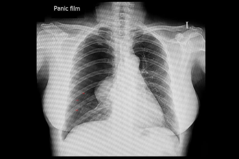
What is a Collapsed Lung (Pneumothorax)?
A hole in the lungs allows air to escape into the space between the lung and chest wall. This accumulation of air and gas in the space can cause the lungs to collapse partially or completely, hence the term 'collapsed lung' is also used.
Pneumothorax often occurs alongside diseases like tuberculosis and is commonly seen in smokers. Another high-risk group includes patients with COPD. This susceptibility is due to their weaker lung structures compared to the general population, making them less capable of preventing spontaneous ruptures.
Types of Pneumothorax
There are mainly two types of pneumothorax: spontaneous and traumatic. Other less common types include tension and menstrual pneumothorax.
Spontaneous Pneumothorax
In spontaneous pneumothorax, the lungs collapse without any external injury. It is divided into primary and secondary spontaneous pneumothorax. In primary spontaneous pneumothorax, the collapse is caused by the rupture of abnormal air pockets in the lungs releasing air. There is no underlying health condition or disease associated with primary spontaneous pneumothorax.
The secondary spontaneous pneumothorax type occurs due to lung diseases which cause obstructions, leading to the formation and bursting of swollen areas known as bullae.
Traumatic Pneumothorax
Another type is traumatic pneumothorax, which is subdivided into two categories. The first type results from injuries such as stab wounds or broken ribs. The other is iatrogenic pneumothorax, which occurs when the lung is punctured during medical procedures like central venous catheter placement or biopsy.
Tension Pneumothorax
This condition occurs when air can enter the lungs but cannot escape, creating a one-way valve effect that leads to increased pressure inside the lungs. Tension pneumothorax is a medical emergency that requires immediate attention.
Menstrual Pneumothorax
Although rare, menstrual pneumothorax affects women with endometriosis. Endometrial tissue, growing outside the uterus, can bleed into the pleural space, creating cysts that may lead to lung collapse.
Causes of Pneumothorax
Pneumothorax can stem from various conditions and diseases. There are primarily two types of causative circumstances.
The first scenario involves people without prior lung disease diagnosis but have conditions such as asthma, cystic fibrosis, COPD, tuberculosis, emphysema, and whooping cough which can complicate into pneumothorax. Pneumothorax occurs in about 70% of people with COPD. Other medical conditions that can cause it include:
- Lung agglomeration
- Collagen vascular disease
- Idiopathic pulmonary fibrosis
- Lung cancer
- Lymphangioleiomyomatosis
- Acute respiratory distress syndrome
The second causative situation is traumatic reasons. Pneumothorax can occur due to lung tissue integrity being compromised by causes such as stab wounds, gunshot wounds, and rib fractures. Additionally, medical procedures such as biopsies or venous catheterization can also lead to traumatic pneumothorax. Other causes within traumatic pneumothorax include:
- Blunt force trauma
- Nerve block
- Mechanical ventilation
Besides these, small bubbles that form between the lung and its outer surface during embryogenesis generally affect young males and can also cause spontaneous pneumothorax.
Another rare cause of pneumothorax is menstrual pneumothorax in women during their menstrual periods. This condition, known as catamenial pneumothorax, occurs when endometrial tissue attaches to the lungs and forms cysts. These cysts allow blood and air to enter the pleural space, leading to lung collapse.
In addition to these causes, certain lifestyle factors can also lead to pneumothorax. These include:
- Drug use
- Flights involving significant changes in air pressure
- Scuba diving
Risk Factors
Factors that increase the risk of pneumothorax include:
- Pneumothorax is seven times more common in men than in women.
- Being tall increases the risk of pneumothorax in men.
- Smoking is considered the biggest risk factor for pneumothorax.
- One in ten people with a family history of pneumothorax will experience it.
- Being pregnant
- Marfan syndrome
Symptoms of Pneumothorax
Symptoms of a collapsed lung can occur during wakeful rest, sleep, or following chest trauma. In cases where the hole is small, no symptoms may be apparent, allowing for self-healing. However, with a larger hole in the lung, symptoms include:
- Rapid heart rate
- Skin discoloration to blue or ash due to lack of oxygen
- Easily fatigued
- Feeling of tightness in the chest
- Shortness of breath or feeling unable to breathe deeply
- Sharp chest pain that can spread to the shoulder, arm, or back, especially when coughing or taking deep breaths
- Swelling of the neck veins
- Flaring of the nostrils
- Low blood pressure
- Anxiety
Due to the varying severity of symptoms among patients, it can take a few days to recognize that something is wrong. However, seeking immediate medical help is crucial upon noticing any signs of a collapsed lung, as it is a life-threatening condition.
How is Pneumothorax Diagnosed?
Physical examination is crucial in the diagnosis of pneumothorax. Specialists can determine the presence of a collapsed lung by listening to the breath sounds on the affected side, which may be diminished or absent. Normally, both sides of the chest should rise and fall equally during breathing. In cases of pneumothorax, one side of the chest may not rise. Other tests to confirm the diagnosis of pneumothorax include:
- Chest X-ray
- Computed Tomography (CT)
- Ultrasound
- Arterial blood gas test
Treatment Methods for Pneumothorax
In cases where the lung tear is small, the condition may resolve on its own, but large tears often require hospitalization. The primary goal of treating a pneumothorax is to remove the air and re-expand the lung. This is typically achieved through a procedure called needle aspiration.
Needle Aspiration
In needle aspiration, a needle is inserted through the rib space into the space between the lung and chest wall. Subsequently, a chest tube may be placed, and it can remain in place for several days until the condition that caused the pneumothorax resolves. If this procedure is not effective, chest surgery may be performed.
Needle aspiration can be a painful process, so regional pain relief or anesthesia may be administered. Additionally, antibiotics may be started to prevent potential infections. Patients treated in the emergency room can be referred to pulmonary specialists or thoracic surgeons for advanced care.
Chemical Pleurodesis
To prevent the lungs from collapsing again, pleurodesis may be performed. This involves the surgeon making an incision and then placing a tube. Various chemicals are then used to seal the hole, attaching the lung to the chest cavity.
Surgical Treatment of Pneumothorax
While the above treatments are often sufficient, surgical procedures may be required in some cases. Surgery is recommended for patients with the following conditions who do not respond to other treatments:
- Traumatic lung injuries
- Collapse of both lungs
- Recurrent lung collapse
- Lung that does not re-expand despite the insertion of a chest tube
- Persistent air leak from the chest tube
Surgical Methods for Treating Pneumothorax
Surgical procedures for treating pneumothorax can be either open or closed.
Closed Method
Surgical intervention for pneumothorax often involves minimally invasive techniques, among which VATS (Video-Assisted Thoracoscopic Surgery) is the most commonly used. This procedure involves entering the chest cavity through a single 2-centimeter incision. A camera and surgical instruments are then used to repair the hole in the lung. This method is favored because it causes minimal damage to healthy tissue, leading to faster and more comfortable recovery for patients.
Known as the endoscopic stapling method, VATS is particularly suitable for patients with the following characteristics:
- Patients with recurrent pneumothorax
- Detection of bullae or air pockets
- Individuals in high-risk occupational groups
The procedure for treating pneumothorax generally lasts about 45 minutes. In cases of atelectasis-type pneumothorax, the procedure is limited to 10-15 minutes. Patients typically need to stay in the hospital for 2-3 days post-surgery.
Open Method
Although rarely preferred today, open surgery is still a method for treating pneumothorax. This procedure involves making a 7-8 centimeter incision between the third and fourth ribs. Air sacs in the lung are then removed through a specialized procedure, and it is also possible to remove part of the lung's outer section. Subsequently, a reaction called pleurodesis is induced to cause the lungs to adhere to the chest cavity.
At the end of the surgery, a drain is placed inside the chest cavity and is anatomically sealed. The drain is usually removed 1-2 days after the surgery.
Complications of Pneumothorax Surgery
Some risky conditions can occur after pneumothorax surgery. The likelihood of complications varies between 2% and 10% in surgeries performed using the closed method. Possible complications after pneumothorax surgery include:
- Air leaks in the lung
- Bleeding
- Infection
- Recurrence of the condition
Life After Pneumothorax Surgery
After treatment for pneumothorax, the first thing patients should avoid is flying or scuba diving, especially within the first 2 weeks. Patients with recurrent pneumothorax must be particularly cautious with these activities.
The risk of experiencing a second pneumothorax after the first one is significantly high. Patients should be very careful especially in the first 30 days, and the risk of recurrence remains high throughout the first year. Studies show that the likelihood of experiencing another pneumothorax within the first three years ranges from 20% to 60%. However, generally, no long-term complications are seen after recovery from pneumothorax.
Other considerations for patients after pneumothorax include:
- Not missing monthly doctor visits.
- Avoiding heavy lifting.
- Staying away from strenuous physical activities and sports for at least 6 months after surgery.
- Engaging in regular walking.
- Avoiding smoking and environments where smoking occurs.
- Paying attention to bowel health and consuming a fiber-rich diet to prevent constipation.
- Taking prescribed medications regularly.
Recovery Process from Pneumothorax
Patients with pneumothorax may need to stay in the hospital for a few days for treatment and monitoring. During this time, doctors closely monitor their condition and may administer oxygen if necessary.
Recovery from pneumothorax generally occurs within 2 weeks as the body reabsorbs the extra air around the lungs, allowing them to re-inflate. Additionally, many individuals with a lung puncture heal without needing further treatment.
Complications of Pneumothorax
While recovery from pneumothorax is typically uneventful, some cases can experience severe complications. These include:
- Heart failure
- Respiratory failure
- Lung edema due to damage or infection caused by the treatment


